 Movie
Movie Controller

[English] 日本語
 Yorodumi
- PDB-5a4p: Structure of UBE2Z provides functional insight into specificity i... -
+ Open data

- Basic information
| Entry | Database: PDB / ID: 5a4p | ||||||
|---|---|---|---|---|---|---|---|
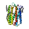
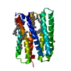
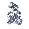
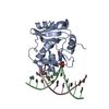
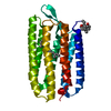
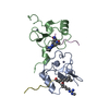
| Title | Structure of UBE2Z provides functional insight into specificity in the FAT10 conjugation machinery | ||||||
 Components Components | UBIQUITIN-CONJUGATING ENZYME E2 Z | ||||||
 Keywords Keywords |  LIGASE / E2 ENZYME / FAT10 CONJUGATION / UBIQUITIN CONJUGATION LIGASE / E2 ENZYME / FAT10 CONJUGATION / UBIQUITIN CONJUGATION | ||||||
| Function / homology |  Function and homology informationE2 ubiquitin-conjugating enzyme / cysteine-type endopeptidase inhibitor activity / ubiquitin conjugating enzyme activity / Synthesis of active ubiquitin: roles of E1 and E2 enzymes / Antigen processing: Ubiquitination & Proteasome degradation / ubiquitin-dependent protein catabolic process / protein ubiquitination / positive regulation of apoptotic process / apoptotic process / negative regulation of apoptotic process ...E2 ubiquitin-conjugating enzyme / cysteine-type endopeptidase inhibitor activity / ubiquitin conjugating enzyme activity / Synthesis of active ubiquitin: roles of E1 and E2 enzymes / Antigen processing: Ubiquitination & Proteasome degradation / ubiquitin-dependent protein catabolic process / protein ubiquitination / positive regulation of apoptotic process / apoptotic process / negative regulation of apoptotic process / nucleoplasm / ATP binding / nucleus / cytosol Function and homology informationE2 ubiquitin-conjugating enzyme / cysteine-type endopeptidase inhibitor activity / ubiquitin conjugating enzyme activity / Synthesis of active ubiquitin: roles of E1 and E2 enzymes / Antigen processing: Ubiquitination & Proteasome degradation / ubiquitin-dependent protein catabolic process / protein ubiquitination / positive regulation of apoptotic process / apoptotic process / negative regulation of apoptotic process ...E2 ubiquitin-conjugating enzyme / cysteine-type endopeptidase inhibitor activity / ubiquitin conjugating enzyme activity / Synthesis of active ubiquitin: roles of E1 and E2 enzymes / Antigen processing: Ubiquitination & Proteasome degradation / ubiquitin-dependent protein catabolic process / protein ubiquitination / positive regulation of apoptotic process / apoptotic process / negative regulation of apoptotic process / nucleoplasm / ATP binding / nucleus / cytosolSimilarity search - Function | ||||||
| Biological species |  HOMO SAPIENS (human) HOMO SAPIENS (human) | ||||||
| Method | X-RAY DIFFRACTION / SYNCHROTRON / MOLECULAR REPLACEMENT / Resolution: 2.1 Å | ||||||
 Authors Authors | Schelpe, J. / Monte, D. / Dewitte, F. / Sixma, T.K. / Rucktooa, P. | ||||||
 Citation Citation | Journal: J.Biol.Chem. / Year: 2016 Title: Structure of Ube2Z Provides Functional Insight Into Specificity in the Fat10 Conjugation Machinery Authors: Schelpe, J. / Monte, D. / Dewitte, F. / Sixma, T.K. / Rucktooa, P. | ||||||
| History |
|
- Structure visualization
| Structure viewer | Molecule: MolmilJmol/JSmol |
|---|
- Downloads & links
-Download
| PDBx/mmCIF format | 5a4p.cif.gz | 108.2 KB | Display | PDBx/mmCIF format |
|---|---|---|---|---|
| PDB format | pdb5a4p.ent.gz | 84.6 KB | Display | PDB format |
| PDBx/mmJSON format | 5a4p.json.gz | Tree view | PDBx/mmJSON format | |
| Others |  Other downloads Other downloads |
-Validation report
| Arichive directory | https://data.pdbj.org/pub/pdb/validation_reports/a4/5a4pftp://data.pdbj.org/pub/pdb/validation_reports/a4/5a4p | HTTPS FTP |
|---|
-Related structure data
| Related structure data |  1x23S S: Starting model for refinement |
|---|---|
| Similar structure data |


-Links
 PDBj
PDBj

- Assembly
| Deposited unit | 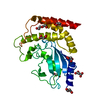
| ||||||||
|---|---|---|---|---|---|---|---|---|---|
| 1 |
| ||||||||
| Unit cell |
|
-Components
| #1: Protein | Mass: 38249.082 Da / Num. of mol.: 1 Source method: isolated from a genetically manipulated source Source: (gene. exp.) HOMO SAPIENS (human) / Plasmid: PETNKI-HIS-3C-LIC-KAN / Production host:  ESCHERICHIA COLI (E. coli) / Strain (production host): BL21(DE3) / References: UniProt: Q9H832, ubiquitin-protein ligase ESCHERICHIA COLI (E. coli) / Strain (production host): BL21(DE3) / References: UniProt: Q9H832, ubiquitin-protein ligase | ||||
|---|---|---|---|---|---|
| #2: Chemical | ChemComp-MLI / Malonic acid  Mass: 102.046 Da / Num. of mol.: 5 / Source method: obtained synthetically / Formula: C3H2O4 Mass: 102.046 Da / Num. of mol.: 5 / Source method: obtained synthetically / Formula: C3H2O4#3: Chemical | Diethylene glycol  Mass: 106.120 Da / Num. of mol.: 2 / Source method: obtained synthetically / Formula: C4H10O3 Mass: 106.120 Da / Num. of mol.: 2 / Source method: obtained synthetically / Formula: C4H10O3#4: Water | ChemComp-HOH / | Water Mass: 18.015 Da / Num. of mol.: 23 / Source method: isolated from a natural source / Formula: H2O Mass: 18.015 Da / Num. of mol.: 23 / Source method: isolated from a natural source / Formula: H2O |
-Experimental details
-Experiment
| Experiment | Method: X-RAY DIFFRACTION / Number of used crystals: 1 |
|---|
- Sample preparation
| Crystal | Density Matthews: 1.79 Å3/Da / Density % sol: 31 % / Description: NONE |
|---|---|
| Crystal grow | Details: 17% (W/V) PEG1500, 0.1M MMT PH 6.5 |
-Data collection
| Diffraction | Mean temperature: 100 K |
|---|---|
| Diffraction source | Source: SYNCHROTRON / Site: ESRF  / Beamline: ID23-1 / Wavelength: 1.03679 / Beamline: ID23-1 / Wavelength: 1.03679 |
| Detector | Type: ADSC QUANTUM 315r / Detector: CCD / Date: Jul 11, 2011 / Details: BENT MIRROR |
| Radiation | Monochromator: SILICON CRYSTAL / Protocol: SINGLE WAVELENGTH / Monochromatic (M) / Laue (L): M / Scattering type: x-ray |
| Radiation wavelength | Wavelength: 1.03679 Å / Relative weight: 1 |
| Reflection | Resolution: 2.1→38.89 Å / Num. obs: 16612 / % possible obs: 94.3 % / Observed criterion σ(I): 0 / Redundancy: 2.7 % / Biso Wilson estimate: 32.33 Å2 / Rmerge(I) obs: 0.1 / Net I/σ(I): 8 |
| Reflection shell | Resolution: 2.1→2.16 Å / Redundancy: 2.7 % / Rmerge(I) obs: 0.59 / Mean I/σ(I) obs: 1.6 / % possible all: 95.2 |
- Processing
| Software |
| ||||||||||||||||||||||||||||||||||||||||||||||||||||||||||||||||||||||||||||||||||||||||||||||||||||||||||||||||||
|---|---|---|---|---|---|---|---|---|---|---|---|---|---|---|---|---|---|---|---|---|---|---|---|---|---|---|---|---|---|---|---|---|---|---|---|---|---|---|---|---|---|---|---|---|---|---|---|---|---|---|---|---|---|---|---|---|---|---|---|---|---|---|---|---|---|---|---|---|---|---|---|---|---|---|---|---|---|---|---|---|---|---|---|---|---|---|---|---|---|---|---|---|---|---|---|---|---|---|---|---|---|---|---|---|---|---|---|---|---|---|---|---|---|---|---|
| Refinement | Method to determine structure: MOLECULAR REPLACEMENT Starting model: PDB ENTRY 1X23 Resolution: 2.1→38.88 Å / Cor.coef. Fo:Fc: 0.915 / Cor.coef. Fo:Fc free: 0.8777 / SU R Cruickshank DPI: 0.227 / Cross valid method: THROUGHOUT / σ(F): 0 / SU R Blow DPI: 0.229 / SU Rfree Blow DPI: 0.197 / SU Rfree Cruickshank DPI: 0.198 Details: IDEAL-DIST CONTACT TERM CONTACT SETUP. ALL ATOMS HAVE CCP4 ATOM TYPE FROM LIBRARY
| ||||||||||||||||||||||||||||||||||||||||||||||||||||||||||||||||||||||||||||||||||||||||||||||||||||||||||||||||||
| Displacement parameters | Biso mean: 58.3 Å2
| ||||||||||||||||||||||||||||||||||||||||||||||||||||||||||||||||||||||||||||||||||||||||||||||||||||||||||||||||||
| Refine analyze | Luzzati coordinate error obs: 0.425 Å | ||||||||||||||||||||||||||||||||||||||||||||||||||||||||||||||||||||||||||||||||||||||||||||||||||||||||||||||||||
| Refinement step | Cycle: LAST / Resolution: 2.1→38.88 Å
| ||||||||||||||||||||||||||||||||||||||||||||||||||||||||||||||||||||||||||||||||||||||||||||||||||||||||||||||||||
| Refine LS restraints |
| ||||||||||||||||||||||||||||||||||||||||||||||||||||||||||||||||||||||||||||||||||||||||||||||||||||||||||||||||||
| LS refinement shell | Resolution: 2.1→2.25 Å / Total num. of bins used: 8
| ||||||||||||||||||||||||||||||||||||||||||||||||||||||||||||||||||||||||||||||||||||||||||||||||||||||||||||||||||
| Refinement TLS params. | Method: refined / Origin x: 18.1054 Å / Origin y: 22.8326 Å / Origin z: 121.425 Å
| ||||||||||||||||||||||||||||||||||||||||||||||||||||||||||||||||||||||||||||||||||||||||||||||||||||||||||||||||||
| Refinement TLS group | Selection details: { A|* } |
