 Movie
Movie Controller

[English] 日本語
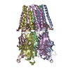
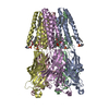
 Yorodumi
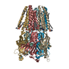
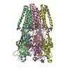
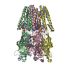
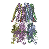
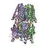
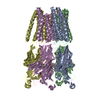
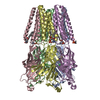
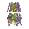
- PDB-4zzc: The GLIC pentameric Ligand-Gated Ion Channel open form complexed ... -
+ Open data

- Basic information
| Entry | Database: PDB / ID: 4zzc | ||||||
|---|---|---|---|---|---|---|---|
| Title | The GLIC pentameric Ligand-Gated Ion Channel open form complexed to xenon | ||||||
 Components Components | Proton-gated ion channel | ||||||
 Keywords Keywords |  MEMBRANE PROTEIN / TRANSPORT PROTEIN MEMBRANE PROTEIN / TRANSPORT PROTEIN | ||||||
| Function / homology |  Function and homology informationsodium channel activity / extracellular ligand-gated monoatomic ion channel activity / transmembrane transporter complex / potassium channel activity / regulation of membrane potential / transmembrane signaling receptor activity / neuron projection / signal transduction / identical protein binding / plasma membrane Function and homology informationsodium channel activity / extracellular ligand-gated monoatomic ion channel activity / transmembrane transporter complex / potassium channel activity / regulation of membrane potential / transmembrane signaling receptor activity / neuron projection / signal transduction / identical protein binding / plasma membraneSimilarity search - Function | ||||||
| Biological species |  Gloeobacter violaceus (bacteria) Gloeobacter violaceus (bacteria) | ||||||
| Method | X-RAY DIFFRACTION / SYNCHROTRON / Resolution: 3.1 Å | ||||||
 Authors Authors | Sauguet, L. / Fourati, Z. / Prange, T. / Delarue, M. / Colloc'h, N. | ||||||
 Citation Citation | Journal: Plos One / Year: 2016 Title: Structural Basis for Xenon Inhibition in a Cationic Pentameric Ligand-Gated Ion Channel. Authors: Sauguet, L. / Fourati, Z. / Prange, T. / Delarue, M. / Colloc'h, N. | ||||||
| History |
|
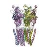
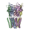
- Structure visualization
| Structure viewer | Molecule: MolmilJmol/JSmol |
|---|
- Downloads & links
-Download
| PDBx/mmCIF format | 4zzc.cif.gz | 648.7 KB | Display | PDBx/mmCIF format |
|---|---|---|---|---|
| PDB format | pdb4zzc.ent.gz | 546.8 KB | Display | PDB format |
| PDBx/mmJSON format | 4zzc.json.gz | Tree view | PDBx/mmJSON format | |
| Others |  Other downloads Other downloads |
-Validation report
| Arichive directory | https://data.pdbj.org/pub/pdb/validation_reports/zz/4zzcftp://data.pdbj.org/pub/pdb/validation_reports/zz/4zzc | HTTPS FTP |
|---|
-Related structure data


-Links
 PDBj
PDBj


- Assembly
| Deposited unit | 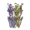
| ||||||||
|---|---|---|---|---|---|---|---|---|---|
| 1 |
| ||||||||
| Unit cell |
|
-Components
-Protein / Sugars , 2 types, 11 molecules ABCDE
| #1: Protein | Mass: 36277.723 Da / Num. of mol.: 5 Source method: isolated from a genetically manipulated source Source: (gene. exp.) Gloeobacter violaceus (strain PCC 7421) (bacteria)Strain: PCC 7421 / Gene: glvI, glr4197 / Production host: Escherichia coli (E. coli) / References: UniProt: Q7NDN8#6: Sugar | ChemComp-LMT /  Type: D-saccharide / Mass: 510.615 Da / Num. of mol.: 6 / Source method: obtained synthetically / Formula: C24H46O11 / Comment: detergent*YM Type: D-saccharide / Mass: 510.615 Da / Num. of mol.: 6 / Source method: obtained synthetically / Formula: C24H46O11 / Comment: detergent*YM |
|---|
-Non-polymers , 6 types, 68 molecules 


| #2: Chemical | ChemComp-ACT / Acetate Mass: 59.044 Da / Num. of mol.: 10 / Source method: obtained synthetically / Formula: C2H3O2 Mass: 59.044 Da / Num. of mol.: 10 / Source method: obtained synthetically / Formula: C2H3O2#3: Chemical | ChemComp-PLC /  Mass: 622.834 Da / Num. of mol.: 10 / Source method: obtained synthetically / Formula: C32H65NO8P / Comment: phospholipid*YM Mass: 622.834 Da / Num. of mol.: 10 / Source method: obtained synthetically / Formula: C32H65NO8P / Comment: phospholipid*YM#4: Chemical | ChemComp-CL / Chloride Mass: 35.453 Da / Num. of mol.: 5 / Source method: obtained synthetically / Formula: Cl Mass: 35.453 Da / Num. of mol.: 5 / Source method: obtained synthetically / Formula: Cl#5: Chemical | ChemComp-NA /  Mass: 22.990 Da / Num. of mol.: 6 / Source method: obtained synthetically / Formula: Na Mass: 22.990 Da / Num. of mol.: 6 / Source method: obtained synthetically / Formula: Na#7: Chemical | ChemComp-XE / Xenon Mass: 131.293 Da / Num. of mol.: 30 / Source method: obtained synthetically / Formula: Xe Mass: 131.293 Da / Num. of mol.: 30 / Source method: obtained synthetically / Formula: Xe#8: Water | ChemComp-HOH / | WaterMass: 18.015 Da / Num. of mol.: 7 / Source method: isolated from a natural source / Formula: H2O |
|---|
-Experimental details
-Experiment
| Experiment | Method: X-RAY DIFFRACTION |
|---|
- Sample preparation
| Crystal | Density Matthews: 5.13 Å3/Da / Density % sol: 76.03 % |
|---|---|
| Crystal grow | Temperature: 290 K / Method: vapor diffusion, hanging drop Details: 12-15% PEG4000 0.1M Na Acetate pH4 0.4M NaSCN 15% glycerol 5% DMSO PH range: 4 |
-Data collection
| Diffraction | Mean temperature: 100 K |
|---|---|
| Diffraction source | Source: SYNCHROTRON / Site: ESRF  / Beamline: BM30A / Wavelength: 1.182 Å / Beamline: BM30A / Wavelength: 1.182 Å |
| Detector | Type: ADSC QUANTUM 315r / Detector: CCD / Date: Jun 20, 2013 |
| Radiation | Protocol: SINGLE WAVELENGTH / Monochromatic (M) / Laue (L): M / Scattering type: x-ray |
| Radiation wavelength | Wavelength: 1.182 Å / Relative weight: 1 |
| Reflection | Resolution: 3.1→25 Å / Biso Wilson estimate: 78.85 Å2 |
| Reflection shell | Resolution: 3.1→3.27 Å / Redundancy: 3.8 % / Rmerge(I) obs: 0.665 / Mean I/σ(I) obs: 2 / % possible all: 99.9 |
- Processing
| Software |
| ||||||||||||||||||||||||||||||||||||||||||||||||||||||||||||||||||||||||||||||||||||||||||||||||||||||||||||||||||||||||||||||||||||||||||||||||||||||
|---|---|---|---|---|---|---|---|---|---|---|---|---|---|---|---|---|---|---|---|---|---|---|---|---|---|---|---|---|---|---|---|---|---|---|---|---|---|---|---|---|---|---|---|---|---|---|---|---|---|---|---|---|---|---|---|---|---|---|---|---|---|---|---|---|---|---|---|---|---|---|---|---|---|---|---|---|---|---|---|---|---|---|---|---|---|---|---|---|---|---|---|---|---|---|---|---|---|---|---|---|---|---|---|---|---|---|---|---|---|---|---|---|---|---|---|---|---|---|---|---|---|---|---|---|---|---|---|---|---|---|---|---|---|---|---|---|---|---|---|---|---|---|---|---|---|---|---|---|---|---|---|
| Refinement | Resolution: 3.1→25 Å / Cor.coef. Fo:Fc: 0.9075 / Cor.coef. Fo:Fc free: 0.8982 / SU R Cruickshank DPI: 0.64 / Cross valid method: THROUGHOUT / σ(F): 0 / SU R Blow DPI: 0.542 / SU Rfree Blow DPI: 0.299 / SU Rfree Cruickshank DPI: 0.315
| ||||||||||||||||||||||||||||||||||||||||||||||||||||||||||||||||||||||||||||||||||||||||||||||||||||||||||||||||||||||||||||||||||||||||||||||||||||||
| Displacement parameters | Biso mean: 88.07 Å2
| ||||||||||||||||||||||||||||||||||||||||||||||||||||||||||||||||||||||||||||||||||||||||||||||||||||||||||||||||||||||||||||||||||||||||||||||||||||||
| Refine analyze | Luzzati coordinate error obs: 0.634 Å | ||||||||||||||||||||||||||||||||||||||||||||||||||||||||||||||||||||||||||||||||||||||||||||||||||||||||||||||||||||||||||||||||||||||||||||||||||||||
| Refinement step | Cycle: LAST / Resolution: 3.1→25 Å
| ||||||||||||||||||||||||||||||||||||||||||||||||||||||||||||||||||||||||||||||||||||||||||||||||||||||||||||||||||||||||||||||||||||||||||||||||||||||
| Refine LS restraints |
| ||||||||||||||||||||||||||||||||||||||||||||||||||||||||||||||||||||||||||||||||||||||||||||||||||||||||||||||||||||||||||||||||||||||||||||||||||||||
| LS refinement shell | Resolution: 3.1→3.18 Å / Total num. of bins used: 20
| ||||||||||||||||||||||||||||||||||||||||||||||||||||||||||||||||||||||||||||||||||||||||||||||||||||||||||||||||||||||||||||||||||||||||||||||||||||||
| Refinement TLS params. | Method: refined / Refine-ID: X-RAY DIFFRACTION
| ||||||||||||||||||||||||||||||||||||||||||||||||||||||||||||||||||||||||||||||||||||||||||||||||||||||||||||||||||||||||||||||||||||||||||||||||||||||
| Refinement TLS group |
|
