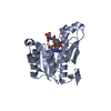
 Movie
Movie Controller

[English] 日本語
 Yorodumi
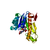
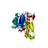
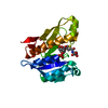
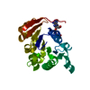
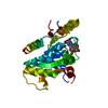
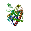
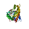
- PDB-4zyw: Human GAR transformylase in complex with GAR and N-({5-[(2-amino-... -
+ Open data

- Basic information
| Entry | Database: PDB / ID: 4zyw | |||||||||
|---|---|---|---|---|---|---|---|---|---|---|
| Title | Human GAR transformylase in complex with GAR and N-({5-[(2-amino-4-oxo-4,7-dihydro-3H-pyrrolo[2,3-d]pyrimidin-6-yl)propyl]thiophen-2-yl}carbonyl)-L-glutamic acid (AGF94) | |||||||||
 Components Components | Trifunctional purine biosynthetic protein adenosine-3 | |||||||||
 Keywords Keywords | TRANSFERASE/TRANSFERASE INHIBITOR /  gar transformylase / antifolate / TRANSFERASE-TRANSFERASE INHIBITOR complex gar transformylase / antifolate / TRANSFERASE-TRANSFERASE INHIBITOR complex | |||||||||
| Function / homology |  Function and homology informationphosphoribosylformylglycinamidine cyclo-ligase / phosphoribosylformylglycinamidine cyclo-ligase activity / adenine biosynthetic process / phosphoribosylamine-glycine ligase / phosphoribosylamine-glycine ligase activity / phosphoribosylglycinamide formyltransferase 1 / purine ribonucleoside monophosphate biosynthetic process / phosphoribosylglycinamide formyltransferase activity / 'de novo' XMP biosynthetic process / brainstem development ...phosphoribosylformylglycinamidine cyclo-ligase / phosphoribosylformylglycinamidine cyclo-ligase activity / adenine biosynthetic process / phosphoribosylamine-glycine ligase / phosphoribosylamine-glycine ligase activity / phosphoribosylglycinamide formyltransferase 1 / purine ribonucleoside monophosphate biosynthetic process / phosphoribosylglycinamide formyltransferase activity / 'de novo' XMP biosynthetic process / brainstem development / Purine ribonucleoside monophosphate biosynthesis / glycine metabolic process / 'de novo' AMP biosynthetic process / GMP biosynthetic process / purine nucleotide biosynthetic process / 'de novo' IMP biosynthetic process / : / tetrahydrofolate biosynthetic process / cerebellum development / : / cerebral cortex development / extracellular exosome / ATP binding / metal ion binding / cytosol Function and homology informationphosphoribosylformylglycinamidine cyclo-ligase / phosphoribosylformylglycinamidine cyclo-ligase activity / adenine biosynthetic process / phosphoribosylamine-glycine ligase / phosphoribosylamine-glycine ligase activity / phosphoribosylglycinamide formyltransferase 1 / purine ribonucleoside monophosphate biosynthetic process / phosphoribosylglycinamide formyltransferase activity / 'de novo' XMP biosynthetic process / brainstem development ...phosphoribosylformylglycinamidine cyclo-ligase / phosphoribosylformylglycinamidine cyclo-ligase activity / adenine biosynthetic process / phosphoribosylamine-glycine ligase / phosphoribosylamine-glycine ligase activity / phosphoribosylglycinamide formyltransferase 1 / purine ribonucleoside monophosphate biosynthetic process / phosphoribosylglycinamide formyltransferase activity / 'de novo' XMP biosynthetic process / brainstem development / Purine ribonucleoside monophosphate biosynthesis / glycine metabolic process / 'de novo' AMP biosynthetic process / GMP biosynthetic process / purine nucleotide biosynthetic process / 'de novo' IMP biosynthetic process / : / tetrahydrofolate biosynthetic process / cerebellum development / : / cerebral cortex development / extracellular exosome / ATP binding / metal ion binding / cytosolSimilarity search - Function | |||||||||
| Biological species |  Homo sapiens (human) Homo sapiens (human) | |||||||||
| Method | X-RAY DIFFRACTION / SYNCHROTRON / MOLECULAR REPLACEMENT / molecular replacement / Resolution: 2.05 Å | |||||||||
 Authors Authors | Deis, S.M. / Dann III, C.E. | |||||||||
| Funding support |  United States, 2items United States, 2items
| |||||||||
 Citation Citation | Journal: Biochemistry / Year: 2016 Title: Structural and Enzymatic Analysis of Tumor-Targeted Antifolates That Inhibit Glycinamide Ribonucleotide Formyltransferase. Authors: Deis, S.M. / Doshi, A. / Hou, Z. / Matherly, L.H. / Gangjee, A. / Dann, C.E. | |||||||||
| History |
|
- Structure visualization
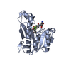
| Structure viewer | Molecule: MolmilJmol/JSmol |
|---|
- Downloads & links
-Download
| PDBx/mmCIF format | 4zyw.cif.gz | 100.5 KB | Display | PDBx/mmCIF format |
|---|---|---|---|---|
| PDB format | pdb4zyw.ent.gz | 74.7 KB | Display | PDB format |
| PDBx/mmJSON format | 4zyw.json.gz | Tree view | PDBx/mmJSON format | |
| Others |  Other downloads Other downloads |
-Validation report
| Arichive directory | https://data.pdbj.org/pub/pdb/validation_reports/zy/4zywftp://data.pdbj.org/pub/pdb/validation_reports/zy/4zyw | HTTPS FTP |
|---|
-Related structure data
| Related structure data | 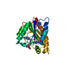 4zytC  4zyuC 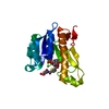 4zyvC 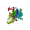 4zyxC  4zyyC 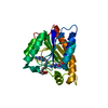 4zyzC  4zz0C  4x72 C: citing same article ( S: Starting model for refinement |
|---|---|
| Similar structure data |


-Links
 PDBj
PDBj


- Assembly
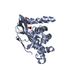
| Deposited unit | 
| ||||||||
|---|---|---|---|---|---|---|---|---|---|
| 1 |
| ||||||||
| Unit cell |
|
-Components
| #1: Protein | Mass: 22810.139 Da / Num. of mol.: 1 / Fragment: gar transformylase domain Source method: isolated from a genetically manipulated source Source: (gene. exp.) Homo sapiens (human) / Gene: GART, PGFT, PRGS / Plasmid: pET22B / Production host:  Escherichia coli (E. coli) / Strain (production host): Rosetta(DE3)pLysS Escherichia coli (E. coli) / Strain (production host): Rosetta(DE3)pLysSReferences: UniProt: P22102, phosphoribosylglycinamide formyltransferase 1 |
|---|---|
| #2: Chemical | ChemComp-GAR / Glycineamide ribonucleotide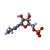  Mass: 284.160 Da / Num. of mol.: 1 / Source method: obtained synthetically / Formula: C7H13N2O8P Mass: 284.160 Da / Num. of mol.: 1 / Source method: obtained synthetically / Formula: C7H13N2O8P |
| #3: Chemical | ChemComp-G94 /   Mass: 447.465 Da / Num. of mol.: 1 / Source method: obtained synthetically / Formula: C19H21N5O6S Mass: 447.465 Da / Num. of mol.: 1 / Source method: obtained synthetically / Formula: C19H21N5O6S |
| #4: Water | ChemComp-HOH / Water Mass: 18.015 Da / Num. of mol.: 104 / Source method: isolated from a natural source / Formula: H2O Mass: 18.015 Da / Num. of mol.: 104 / Source method: isolated from a natural source / Formula: H2O |
-Experimental details
-Experiment
| Experiment | Method: X-RAY DIFFRACTION / Number of used crystals: 1 |
|---|
- Sample preparation
| Crystal | Density Matthews: 3.86 Å3/Da / Density % sol: 68.17 % |
|---|---|
| Crystal grow | Temperature: 277 K / Method: vapor diffusion, sitting drop / pH: 7.5 / Details: 18 % PEG4000, 2 % PEG400, 0.33 M NaCl |
-Data collection
| Diffraction | Mean temperature: 100 K | |||||||||||||||||||||||||||||||||||||||||||||||||||||||||||||||||||||||||||||||||||||||||||||||||||||||||||||||||||||||||||||||||||||||||||||||||||||||||||||||||||||||||||||||||||||||||||||
|---|---|---|---|---|---|---|---|---|---|---|---|---|---|---|---|---|---|---|---|---|---|---|---|---|---|---|---|---|---|---|---|---|---|---|---|---|---|---|---|---|---|---|---|---|---|---|---|---|---|---|---|---|---|---|---|---|---|---|---|---|---|---|---|---|---|---|---|---|---|---|---|---|---|---|---|---|---|---|---|---|---|---|---|---|---|---|---|---|---|---|---|---|---|---|---|---|---|---|---|---|---|---|---|---|---|---|---|---|---|---|---|---|---|---|---|---|---|---|---|---|---|---|---|---|---|---|---|---|---|---|---|---|---|---|---|---|---|---|---|---|---|---|---|---|---|---|---|---|---|---|---|---|---|---|---|---|---|---|---|---|---|---|---|---|---|---|---|---|---|---|---|---|---|---|---|---|---|---|---|---|---|---|---|---|---|---|---|---|---|---|
| Diffraction source | Source: SYNCHROTRON / Site: ALS / Beamline: 4.2.2 / Wavelength: 1.0001 Å | |||||||||||||||||||||||||||||||||||||||||||||||||||||||||||||||||||||||||||||||||||||||||||||||||||||||||||||||||||||||||||||||||||||||||||||||||||||||||||||||||||||||||||||||||||||||||||||
| Detector | Type: RDI CMOS_8M / Detector: CMOS / Date: Apr 6, 2014 | |||||||||||||||||||||||||||||||||||||||||||||||||||||||||||||||||||||||||||||||||||||||||||||||||||||||||||||||||||||||||||||||||||||||||||||||||||||||||||||||||||||||||||||||||||||||||||||
| Radiation | Monochromator: Rosenbaum-Rock Si(111) sagitally focused monochromator Protocol: SINGLE WAVELENGTH / Monochromatic (M) / Laue (L): M / Scattering type: x-ray | |||||||||||||||||||||||||||||||||||||||||||||||||||||||||||||||||||||||||||||||||||||||||||||||||||||||||||||||||||||||||||||||||||||||||||||||||||||||||||||||||||||||||||||||||||||||||||||
| Radiation wavelength | Wavelength: 1.0001 Å / Relative weight: 1 | |||||||||||||||||||||||||||||||||||||||||||||||||||||||||||||||||||||||||||||||||||||||||||||||||||||||||||||||||||||||||||||||||||||||||||||||||||||||||||||||||||||||||||||||||||||||||||||
| Reflection | Resolution: 2.05→50 Å / Num. obs: 21298 / % possible obs: 99.7 % / Redundancy: 10.4 % / Biso Wilson estimate: 24.46 Å2 / Rmerge(I) obs: 0.103 / Rpim(I) all: 0.033 / Rrim(I) all: 0.108 / Χ2: 0.938 / Net I/av σ(I): 23.162 / Net I/σ(I): 10.2 / Num. measured all: 220599 | |||||||||||||||||||||||||||||||||||||||||||||||||||||||||||||||||||||||||||||||||||||||||||||||||||||||||||||||||||||||||||||||||||||||||||||||||||||||||||||||||||||||||||||||||||||||||||||
| Reflection shell | Diffraction-ID: 1 / Rejects: 0
|
-Phasing
| Phasing | Method: molecular replacement |
|---|
- Processing
| Software |
| |||||||||||||||||||||||||||||||||||||||||||||||||||||||||||||||||||||||||||||||||||||||||||||||||||||||||||||||||||||||||||||||||||||||||||||||||||||||||||||||||||||||||||||||||||||||||||||||||||||||||||||||||||||||||||||||||
|---|---|---|---|---|---|---|---|---|---|---|---|---|---|---|---|---|---|---|---|---|---|---|---|---|---|---|---|---|---|---|---|---|---|---|---|---|---|---|---|---|---|---|---|---|---|---|---|---|---|---|---|---|---|---|---|---|---|---|---|---|---|---|---|---|---|---|---|---|---|---|---|---|---|---|---|---|---|---|---|---|---|---|---|---|---|---|---|---|---|---|---|---|---|---|---|---|---|---|---|---|---|---|---|---|---|---|---|---|---|---|---|---|---|---|---|---|---|---|---|---|---|---|---|---|---|---|---|---|---|---|---|---|---|---|---|---|---|---|---|---|---|---|---|---|---|---|---|---|---|---|---|---|---|---|---|---|---|---|---|---|---|---|---|---|---|---|---|---|---|---|---|---|---|---|---|---|---|---|---|---|---|---|---|---|---|---|---|---|---|---|---|---|---|---|---|---|---|---|---|---|---|---|---|---|---|---|---|---|---|---|---|---|---|---|---|---|---|---|---|---|---|---|---|---|---|---|
| Refinement | Method to determine structure: MOLECULAR REPLACEMENT Starting model: 4X72 4x72 Resolution: 2.05→39.825 Å / SU ML: 0.18 / Cross valid method: FREE R-VALUE / σ(F): 1.34 / Phase error: 18.44 / Stereochemistry target values: ML
| |||||||||||||||||||||||||||||||||||||||||||||||||||||||||||||||||||||||||||||||||||||||||||||||||||||||||||||||||||||||||||||||||||||||||||||||||||||||||||||||||||||||||||||||||||||||||||||||||||||||||||||||||||||||||||||||||
| Solvent computation | Shrinkage radii: 0.9 Å / VDW probe radii: 1.11 Å / Solvent model: FLAT BULK SOLVENT MODEL | |||||||||||||||||||||||||||||||||||||||||||||||||||||||||||||||||||||||||||||||||||||||||||||||||||||||||||||||||||||||||||||||||||||||||||||||||||||||||||||||||||||||||||||||||||||||||||||||||||||||||||||||||||||||||||||||||
| Displacement parameters | Biso max: 100.07 Å2 / Biso mean: 35.249 Å2 / Biso min: 11.97 Å2 | |||||||||||||||||||||||||||||||||||||||||||||||||||||||||||||||||||||||||||||||||||||||||||||||||||||||||||||||||||||||||||||||||||||||||||||||||||||||||||||||||||||||||||||||||||||||||||||||||||||||||||||||||||||||||||||||||
| Refinement step | Cycle: final / Resolution: 2.05→39.825 Å
| |||||||||||||||||||||||||||||||||||||||||||||||||||||||||||||||||||||||||||||||||||||||||||||||||||||||||||||||||||||||||||||||||||||||||||||||||||||||||||||||||||||||||||||||||||||||||||||||||||||||||||||||||||||||||||||||||
| Refine LS restraints |
| |||||||||||||||||||||||||||||||||||||||||||||||||||||||||||||||||||||||||||||||||||||||||||||||||||||||||||||||||||||||||||||||||||||||||||||||||||||||||||||||||||||||||||||||||||||||||||||||||||||||||||||||||||||||||||||||||
| LS refinement shell | Refine-ID: X-RAY DIFFRACTION / Total num. of bins used: 14
| |||||||||||||||||||||||||||||||||||||||||||||||||||||||||||||||||||||||||||||||||||||||||||||||||||||||||||||||||||||||||||||||||||||||||||||||||||||||||||||||||||||||||||||||||||||||||||||||||||||||||||||||||||||||||||||||||
| Refinement TLS params. | Method: refined / Refine-ID: X-RAY DIFFRACTION
| |||||||||||||||||||||||||||||||||||||||||||||||||||||||||||||||||||||||||||||||||||||||||||||||||||||||||||||||||||||||||||||||||||||||||||||||||||||||||||||||||||||||||||||||||||||||||||||||||||||||||||||||||||||||||||||||||
| Refinement TLS group |
|
