 Movie
Movie Controller

[English] 日本語
 Yorodumi
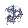
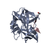
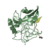
- PDB-4zbx: Family 4 uracil-DNA glycosylase from Sulfolobus tokodaii (free fo... -
+ Open data

- Basic information
| Entry | Database: PDB / ID: 4zbx | ||||||
|---|---|---|---|---|---|---|---|
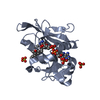
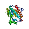
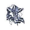
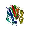
| Title | Family 4 uracil-DNA glycosylase from Sulfolobus tokodaii (free form, X-ray wavelength=0.9000) | ||||||
 Components Components | Uracil-DNA glycosylase | ||||||
 Keywords Keywords | HYDROLASE / Uracil-DNA glycosylase / DNA repair | ||||||
| Function / homology |  Function and homology informationuracil-DNA glycosylase / uracil DNA N-glycosylase activity / 4 iron, 4 sulfur cluster binding / DNA repair / metal ion binding Function and homology informationuracil-DNA glycosylase / uracil DNA N-glycosylase activity / 4 iron, 4 sulfur cluster binding / DNA repair / metal ion bindingSimilarity search - Function | ||||||
| Biological species |   Sulfolobus tokodaii str. 7 (archaea) Sulfolobus tokodaii str. 7 (archaea) | ||||||
| Method | X-RAY DIFFRACTION / SYNCHROTRON / MOLECULAR REPLACEMENT / Resolution: 1.7 Å | ||||||
 Authors Authors | Kawai, A. / Miyamoto, S. | ||||||
 Citation Citation | Journal: Febs Lett. / Year: 2015 Title: Crystal structure of family 4 uracil-DNA glycosylase from Sulfolobus tokodaii and a function of tyrosine 170 in DNA binding Authors: Kawai, A. / Higuchi, S. / Tsunoda, M. / Nakamura, K.T. / Yamagata, Y. / Miyamoto, S. | ||||||
| History |
|
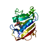
- Structure visualization
| Structure viewer | Molecule: MolmilJmol/JSmol |
|---|
- Downloads & links
-Download
| PDBx/mmCIF format | 4zbx.cif.gz | 101.3 KB | Display | PDBx/mmCIF format |
|---|---|---|---|---|
| PDB format | pdb4zbx.ent.gz | 75.8 KB | Display | PDB format |
| PDBx/mmJSON format | 4zbx.json.gz | Tree view | PDBx/mmJSON format | |
| Others |  Other downloads Other downloads |
-Validation report
| Arichive directory | https://data.pdbj.org/pub/pdb/validation_reports/zb/4zbxftp://data.pdbj.org/pub/pdb/validation_reports/zb/4zbx | HTTPS FTP |
|---|
-Related structure data
| Related structure data |  4zbyC  4zbzC  1ui0S C: citing same article ( S: Starting model for refinement |
|---|---|
| Similar structure data |


-Links
 PDBj
PDBj- Assembly
| Deposited unit | 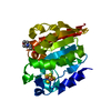
| ||||||||
|---|---|---|---|---|---|---|---|---|---|
| 1 |
| ||||||||
| Unit cell |
|
-Components
| #1: Protein | Mass: 22360.035 Da / Num. of mol.: 1 / Fragment: RESIDUES 1-194 Source method: isolated from a genetically manipulated source Source: (gene. exp.) Sulfolobus tokodaii str. 7 (archaea) / Strain: 7 / Gene: STK_22380 / Plasmid: pET11a / Production host:  Escherichia coli BL21(DE3) (bacteria) / Strain (production host): BL21(DE3) / References: UniProt: Q96YD0, uracil-DNA glycosylase Escherichia coli BL21(DE3) (bacteria) / Strain (production host): BL21(DE3) / References: UniProt: Q96YD0, uracil-DNA glycosylase |
|---|---|
| #2: Chemical | ChemComp-SF4 / Iron–sulfur cluster  Mass: 351.640 Da / Num. of mol.: 1 / Source method: obtained synthetically / Formula: Fe4S4 Mass: 351.640 Da / Num. of mol.: 1 / Source method: obtained synthetically / Formula: Fe4S4 |
| #3: Chemical | ChemComp-MES / MES (buffer)  Mass: 195.237 Da / Num. of mol.: 1 / Source method: obtained synthetically / Formula: C6H13NO4S / Comment: pH buffer*YM Mass: 195.237 Da / Num. of mol.: 1 / Source method: obtained synthetically / Formula: C6H13NO4S / Comment: pH buffer*YM |
| #4: Water | ChemComp-HOH / Water Mass: 18.015 Da / Num. of mol.: 150 / Source method: isolated from a natural source / Formula: H2O Mass: 18.015 Da / Num. of mol.: 150 / Source method: isolated from a natural source / Formula: H2O |
-Experimental details
-Experiment
| Experiment | Method: X-RAY DIFFRACTION |
|---|
- Sample preparation
| Crystal | Density Matthews: 2.3 Å3/Da / Density % sol: 46.51 % |
|---|---|
| Crystal grow | Temperature: 293 K / Method: vapor diffusion, hanging drop / pH: 5.8 / Details: 20% PEG3350, 0.1M MES, 20% glycerol |
-Data collection
| Diffraction | Mean temperature: 100 K |
|---|---|
| Diffraction source | Source: SYNCHROTRON / Site: SPring-8  / Beamline: BL44XU / Wavelength: 0.9 Å / Beamline: BL44XU / Wavelength: 0.9 Å |
| Detector | Type: Bruker DIP-6040 / Detector: IMAGE PLATE / Date: Sep 28, 2007 |
| Radiation | Protocol: SINGLE WAVELENGTH / Monochromatic (M) / Laue (L): M / Scattering type: x-ray |
| Radiation wavelength | Wavelength: 0.9 Å / Relative weight: 1 |
| Reflection | Resolution: 1.7→50 Å / Num. obs: 23097 / % possible obs: 100 % / Redundancy: 7.3 % / Rmerge(I) obs: 0.05 / Net I/σ(I): 50.8 |
| Reflection shell | Resolution: 1.7→1.76 Å / Redundancy: 7.1 % / Rmerge(I) obs: 0.339 / Mean I/σ(I) obs: 6.4 / % possible all: 100 |
- Processing
| Software |
| |||||||||||||||||||||||||||||||||||||||||||||||||||||||||||||||
|---|---|---|---|---|---|---|---|---|---|---|---|---|---|---|---|---|---|---|---|---|---|---|---|---|---|---|---|---|---|---|---|---|---|---|---|---|---|---|---|---|---|---|---|---|---|---|---|---|---|---|---|---|---|---|---|---|---|---|---|---|---|---|---|---|
| Refinement | Method to determine structure: MOLECULAR REPLACEMENT Starting model: 1ui0 Resolution: 1.7→36.925 Å / SU ML: 0.14 / Cross valid method: FREE R-VALUE / σ(F): 1.93 / Phase error: 19.81 / Stereochemistry target values: ML
| |||||||||||||||||||||||||||||||||||||||||||||||||||||||||||||||
| Solvent computation | Shrinkage radii: 0.9 Å / VDW probe radii: 1.11 Å / Solvent model: FLAT BULK SOLVENT MODEL | |||||||||||||||||||||||||||||||||||||||||||||||||||||||||||||||
| Refinement step | Cycle: LAST / Resolution: 1.7→36.925 Å
| |||||||||||||||||||||||||||||||||||||||||||||||||||||||||||||||
| Refine LS restraints |
| |||||||||||||||||||||||||||||||||||||||||||||||||||||||||||||||
| LS refinement shell |
| |||||||||||||||||||||||||||||||||||||||||||||||||||||||||||||||
| Refinement TLS params. | Method: refined / Origin x: -25.047 Å / Origin y: 0.297 Å / Origin z: 8.4553 Å
| |||||||||||||||||||||||||||||||||||||||||||||||||||||||||||||||
| Refinement TLS group | Selection details: (chain 'A' and resid 1 through 192) |
