 Movie
Movie Controller

[English] 日本語
 Yorodumi
- PDB-4zb6: Crystal structure of glutathione transferase URE2P4 from Phaneroc... -
+ Open data

- Basic information
| Entry | Database: PDB / ID: 4zb6 | ||||||
|---|---|---|---|---|---|---|---|
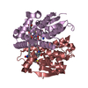
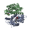
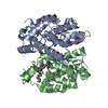
| Title | Crystal structure of glutathione transferase URE2P4 from Phanerochaete chrysosporium in complex with oxidized glutathione. | ||||||
 Components Components | PcUre2p4 | ||||||
 Keywords Keywords |  TRANSFERASE / GLUTATHIONE TRANSFERASE / GST FOLD / OXYDIZED GLUTATHIONE TRANSFERASE / GLUTATHIONE TRANSFERASE / GST FOLD / OXYDIZED GLUTATHIONE | ||||||
| Function / homology |  Function and homology informationGlutathione S-transferase, C-terminal domain / Glutathione S-transferase, N-terminal domain / Glutathione transferase family / Glutathione S-transferase Yfyf (Class Pi); Chain A, domain 2 - #10 / Glutathione S-transferase, C-terminal / Glutathione S-transferase Yfyf (Class Pi); Chain A, domain 2 / Soluble glutathione S-transferase N-terminal domain profile. / Glutathione S-transferase, C-terminal-like / Soluble glutathione S-transferase C-terminal domain profile. / Glutathione S-transferase, N-terminal ...Glutathione S-transferase, C-terminal domain / Glutathione S-transferase, N-terminal domain / Glutathione transferase family / Glutathione S-transferase Yfyf (Class Pi); Chain A, domain 2 - #10 / Glutathione S-transferase, C-terminal / Glutathione S-transferase Yfyf (Class Pi); Chain A, domain 2 / Soluble glutathione S-transferase N-terminal domain profile. / Glutathione S-transferase, C-terminal-like / Soluble glutathione S-transferase C-terminal domain profile. / Glutathione S-transferase, N-terminal / Glutathione S-transferase, C-terminal domain superfamily / Glutaredoxin / Glutaredoxin / Thioredoxin-like superfamily / Up-down Bundle / 3-Layer(aba) Sandwich / Mainly Alpha / Alpha Beta Function and homology informationGlutathione S-transferase, C-terminal domain / Glutathione S-transferase, N-terminal domain / Glutathione transferase family / Glutathione S-transferase Yfyf (Class Pi); Chain A, domain 2 - #10 / Glutathione S-transferase, C-terminal / Glutathione S-transferase Yfyf (Class Pi); Chain A, domain 2 / Soluble glutathione S-transferase N-terminal domain profile. / Glutathione S-transferase, C-terminal-like / Soluble glutathione S-transferase C-terminal domain profile. / Glutathione S-transferase, N-terminal ...Glutathione S-transferase, C-terminal domain / Glutathione S-transferase, N-terminal domain / Glutathione transferase family / Glutathione S-transferase Yfyf (Class Pi); Chain A, domain 2 - #10 / Glutathione S-transferase, C-terminal / Glutathione S-transferase Yfyf (Class Pi); Chain A, domain 2 / Soluble glutathione S-transferase N-terminal domain profile. / Glutathione S-transferase, C-terminal-like / Soluble glutathione S-transferase C-terminal domain profile. / Glutathione S-transferase, N-terminal / Glutathione S-transferase, C-terminal domain superfamily / Glutaredoxin / Glutaredoxin / Thioredoxin-like superfamily / Up-down Bundle / 3-Layer(aba) Sandwich / Mainly Alpha / Alpha BetaSimilarity search - Domain/homology | ||||||
| Biological species |  Phanerochaete chrysosporium (fungus) Phanerochaete chrysosporium (fungus) | ||||||
| Method | X-RAY DIFFRACTION / SYNCHROTRON / MOLECULAR REPLACEMENT / Resolution: 1.801 Å | ||||||
 Authors Authors | Roret, T. / Didierjean, C. | ||||||
 Citation Citation | Journal: Fungal Genet. Biol. / Year: 2015 Title: Evolutionary divergence of Ure2pA glutathione transferases in wood degrading fungi. Authors: Roret, T. / Thuillier, A. / Favier, F. / Gelhaye, E. / Didierjean, C. / Morel-Rouhier, M. | ||||||
| History |
|
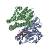
- Structure visualization
| Structure viewer | Molecule: MolmilJmol/JSmol |
|---|
- Downloads & links
-Download
| PDBx/mmCIF format | 4zb6.cif.gz | 382.5 KB | Display | PDBx/mmCIF format |
|---|---|---|---|---|
| PDB format | pdb4zb6.ent.gz | 313.2 KB | Display | PDB format |
| PDBx/mmJSON format | 4zb6.json.gz | Tree view | PDBx/mmJSON format | |
| Others |  Other downloads Other downloads |
-Validation report
| Arichive directory | https://data.pdbj.org/pub/pdb/validation_reports/zb/4zb6ftp://data.pdbj.org/pub/pdb/validation_reports/zb/4zb6 | HTTPS FTP |
|---|
-Related structure data
| Related structure data |  4f0cC  4zb7C  4zb8SC 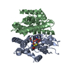 4zb9C 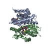 4zbaC  4zbbC  4zbdC S: Starting model for refinement C: citing same article ( |
|---|---|
| Similar structure data |


-Links
 PDBj
PDBj

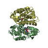
- Assembly
| Deposited unit | 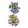
| ||||||||
|---|---|---|---|---|---|---|---|---|---|
| 1 | 
| ||||||||
| 2 | 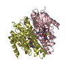
| ||||||||
| Unit cell |
|
-Components
| #1: Protein | Mass: 25647.922 Da / Num. of mol.: 4 Source method: isolated from a genetically manipulated source Source: (gene. exp.) Phanerochaete chrysosporium (fungus) / Production host:  Escherichia coli (E. coli) / References: UniProt: A0A0R4I980*PLUS Escherichia coli (E. coli) / References: UniProt: A0A0R4I980*PLUS#2: Chemical | ChemComp-GDS / Glutathione disulfide  Mass: 612.631 Da / Num. of mol.: 4 / Source method: obtained synthetically / Formula: C20H32N6O12S2 Mass: 612.631 Da / Num. of mol.: 4 / Source method: obtained synthetically / Formula: C20H32N6O12S2#3: Chemical | ChemComp-NA /   Mass: 22.990 Da / Num. of mol.: 4 / Source method: obtained synthetically / Formula: Na Mass: 22.990 Da / Num. of mol.: 4 / Source method: obtained synthetically / Formula: Na#4: Water | ChemComp-HOH / | Water Mass: 18.015 Da / Num. of mol.: 1123 / Source method: isolated from a natural source / Formula: H2O Mass: 18.015 Da / Num. of mol.: 1123 / Source method: isolated from a natural source / Formula: H2O |
|---|
-Experimental details
-Experiment
| Experiment | Method: X-RAY DIFFRACTION |
|---|
- Sample preparation
| Crystal | Density Matthews: 2.22 Å3/Da / Density % sol: 44.51 % |
|---|---|
| Crystal grow | Temperature: 278 K / Method: microbatch Details: 30 % PEG4000, 0.1M Sodium Acetate pH4.6, 0.2M Ammonium Acetate |
-Data collection
| Diffraction | Mean temperature: 100 K |
|---|---|
| Diffraction source | Source: SYNCHROTRON / Site: ESRF  / Beamline: BM30A / Wavelength: 0.999 Å / Beamline: BM30A / Wavelength: 0.999 Å |
| Detector | Type: ADSC QUANTUM 315r / Detector: CCD / Date: Feb 1, 2013 |
| Radiation | Protocol: SINGLE WAVELENGTH / Monochromatic (M) / Laue (L): M / Scattering type: x-ray |
| Radiation wavelength | Wavelength: 0.999 Å / Relative weight: 1 |
| Reflection | Resolution: 1.8→47 Å / Num. obs: 84906 / % possible obs: 99.9 % / Redundancy: 7.3 % / Net I/σ(I): 14.5 |
| Reflection shell | Resolution: 1.8→1.9 Å / Redundancy: 7.1 % / Rmerge(I) obs: 0.761 / Mean I/σ(I) obs: 3.3 / Num. unique all: 12133 / % possible all: 99.1 |
- Processing
| Software |
| |||||||||||||||||||||||||||||||||||||||||||||||||||||||||||||||||||||||||||||||||||||||||||||||||||||||||||||||||||||||||||||||||||||||||||||||||||||||||||||||||||||||||||||||||||||||||||||||||||||||||||||||||||||||||
|---|---|---|---|---|---|---|---|---|---|---|---|---|---|---|---|---|---|---|---|---|---|---|---|---|---|---|---|---|---|---|---|---|---|---|---|---|---|---|---|---|---|---|---|---|---|---|---|---|---|---|---|---|---|---|---|---|---|---|---|---|---|---|---|---|---|---|---|---|---|---|---|---|---|---|---|---|---|---|---|---|---|---|---|---|---|---|---|---|---|---|---|---|---|---|---|---|---|---|---|---|---|---|---|---|---|---|---|---|---|---|---|---|---|---|---|---|---|---|---|---|---|---|---|---|---|---|---|---|---|---|---|---|---|---|---|---|---|---|---|---|---|---|---|---|---|---|---|---|---|---|---|---|---|---|---|---|---|---|---|---|---|---|---|---|---|---|---|---|---|---|---|---|---|---|---|---|---|---|---|---|---|---|---|---|---|---|---|---|---|---|---|---|---|---|---|---|---|---|---|---|---|---|---|---|---|---|---|---|---|---|---|---|---|---|---|---|---|---|
| Refinement | Method to determine structure: MOLECULAR REPLACEMENT Starting model: 4ZB8 Resolution: 1.801→47 Å / SU ML: 0.19 / Cross valid method: FREE R-VALUE / σ(F): 1.35 / Phase error: 18.05 / Stereochemistry target values: ML
| |||||||||||||||||||||||||||||||||||||||||||||||||||||||||||||||||||||||||||||||||||||||||||||||||||||||||||||||||||||||||||||||||||||||||||||||||||||||||||||||||||||||||||||||||||||||||||||||||||||||||||||||||||||||||
| Solvent computation | Shrinkage radii: 0.73 Å / VDW probe radii: 1 Å / Solvent model: FLAT BULK SOLVENT MODEL / Bsol: 40.862 Å2 / ksol: 0.361 e/Å3 | |||||||||||||||||||||||||||||||||||||||||||||||||||||||||||||||||||||||||||||||||||||||||||||||||||||||||||||||||||||||||||||||||||||||||||||||||||||||||||||||||||||||||||||||||||||||||||||||||||||||||||||||||||||||||
| Displacement parameters |
| |||||||||||||||||||||||||||||||||||||||||||||||||||||||||||||||||||||||||||||||||||||||||||||||||||||||||||||||||||||||||||||||||||||||||||||||||||||||||||||||||||||||||||||||||||||||||||||||||||||||||||||||||||||||||
| Refinement step | Cycle: LAST / Resolution: 1.801→47 Å
| |||||||||||||||||||||||||||||||||||||||||||||||||||||||||||||||||||||||||||||||||||||||||||||||||||||||||||||||||||||||||||||||||||||||||||||||||||||||||||||||||||||||||||||||||||||||||||||||||||||||||||||||||||||||||
| Refine LS restraints |
| |||||||||||||||||||||||||||||||||||||||||||||||||||||||||||||||||||||||||||||||||||||||||||||||||||||||||||||||||||||||||||||||||||||||||||||||||||||||||||||||||||||||||||||||||||||||||||||||||||||||||||||||||||||||||
| LS refinement shell |
|
