 Movie
Movie Controller

[English] 日本語
 Yorodumi
- PDB-4zaj: 2.2 Angstrom Crystal Structure of a Human Arginyl-tRNA Synthetase -
+ Open data

- Basic information
| Entry | Database: PDB / ID: 4zaj | ||||||
|---|---|---|---|---|---|---|---|
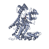
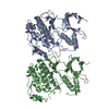
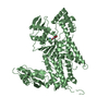
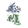
| Title | 2.2 Angstrom Crystal Structure of a Human Arginyl-tRNA Synthetase | ||||||
 Components Components | Arginine--tRNA ligase, cytoplasmic | ||||||
 Keywords Keywords |  LIGASE / tRNA arginine synthetase Arg-tRNA LIGASE / tRNA arginine synthetase Arg-tRNA | ||||||
| Function / homology |  Function and homology informationarginine-tRNA ligase / arginine-tRNA ligase activity / arginyl-tRNA aminoacylation / Selenoamino acid metabolism / Cytosolic tRNA aminoacylation / aminoacyl-tRNA synthetase multienzyme complex / tRNA aminoacylation for protein translation / arginine binding / tRNA binding / cadherin binding ...arginine-tRNA ligase / arginine-tRNA ligase activity / arginyl-tRNA aminoacylation / Selenoamino acid metabolism / Cytosolic tRNA aminoacylation / aminoacyl-tRNA synthetase multienzyme complex / tRNA aminoacylation for protein translation / arginine binding / tRNA binding / cadherin binding / nucleolus / extracellular exosome / nucleoplasm / ATP binding / membrane / nucleus / cytosol / cytoplasm Function and homology informationarginine-tRNA ligase / arginine-tRNA ligase activity / arginyl-tRNA aminoacylation / Selenoamino acid metabolism / Cytosolic tRNA aminoacylation / aminoacyl-tRNA synthetase multienzyme complex / tRNA aminoacylation for protein translation / arginine binding / tRNA binding / cadherin binding ...arginine-tRNA ligase / arginine-tRNA ligase activity / arginyl-tRNA aminoacylation / Selenoamino acid metabolism / Cytosolic tRNA aminoacylation / aminoacyl-tRNA synthetase multienzyme complex / tRNA aminoacylation for protein translation / arginine binding / tRNA binding / cadherin binding / nucleolus / extracellular exosome / nucleoplasm / ATP binding / membrane / nucleus / cytosol / cytoplasmSimilarity search - Function | ||||||
| Biological species |  Homo sapiens (human) Homo sapiens (human) | ||||||
| Method | X-RAY DIFFRACTION / SYNCHROTRON / MOLECULAR REPLACEMENT / molecular replacement / Resolution: 2.22 Å | ||||||
 Authors Authors | Smith, A.T. / Rosenzweig, A.C. | ||||||
 Citation Citation | Journal: To Be Published Title: 2.2 Angstrom crystal structure of a human Arginyl-tRNA synthetase Authors: Smith, A.T. / Rosenzweig, A.C. | ||||||
| History |
|
- Structure visualization
| Structure viewer | Molecule: MolmilJmol/JSmol |
|---|
- Downloads & links
-Download
| PDBx/mmCIF format | 4zaj.cif.gz | 128.3 KB | Display | PDBx/mmCIF format |
|---|---|---|---|---|
| PDB format | pdb4zaj.ent.gz | 99.5 KB | Display | PDB format |
| PDBx/mmJSON format | 4zaj.json.gz | Tree view | PDBx/mmJSON format | |
| Others |  Other downloads Other downloads |
-Validation report
| Arichive directory | https://data.pdbj.org/pub/pdb/validation_reports/za/4zajftp://data.pdbj.org/pub/pdb/validation_reports/za/4zaj | HTTPS FTP |
|---|
-Related structure data
| Related structure data |  4q2tS S: Starting model for refinement |
|---|---|
| Similar structure data |


-Links
 PDBj
PDBj
- Assembly
| Deposited unit | 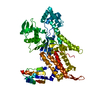
| ||||||||
|---|---|---|---|---|---|---|---|---|---|
| 1 |
| ||||||||
| Unit cell |
| ||||||||
| Details | Monomer by size exclusion chromatography |
-Components
| #1: Protein | Mass: 68941.180 Da / Num. of mol.: 1 Source method: isolated from a genetically manipulated source Source: (gene. exp.) Homo sapiens (human) / Gene: RARS / Production host:  Escherichia coli (E. coli) / References: UniProt: P54136, arginine-tRNA ligase Escherichia coli (E. coli) / References: UniProt: P54136, arginine-tRNA ligase |
|---|---|
| #2: Water | ChemComp-HOH / Water Mass: 18.015 Da / Num. of mol.: 106 / Source method: isolated from a natural source / Formula: H2O Mass: 18.015 Da / Num. of mol.: 106 / Source method: isolated from a natural source / Formula: H2O |
-Experimental details
-Experiment
| Experiment | Method: X-RAY DIFFRACTION / Number of used crystals: 1 |
|---|
- Sample preparation
| Crystal | Density Matthews: 2.63 Å3/Da / Density % sol: 53.17 % |
|---|---|
| Crystal grow | Temperature: 293 K / Method: vapor diffusion, sitting drop / pH: 6.5 / Details: 25% PEG 1000 / Temp details: ambient temperature |
-Data collection
| Diffraction | Mean temperature: 100 K | ||||||||||||||||||||||||
|---|---|---|---|---|---|---|---|---|---|---|---|---|---|---|---|---|---|---|---|---|---|---|---|---|---|
| Diffraction source | Source: SYNCHROTRON / Site: APS  / Beamline: 21-ID-G / Wavelength: 0.97857 Å / Beamline: 21-ID-G / Wavelength: 0.97857 Å | ||||||||||||||||||||||||
| Detector | Type: MAR scanner 300 mm plate / Detector: IMAGE PLATE / Date: Mar 26, 2015 | ||||||||||||||||||||||||
| Radiation | Protocol: SINGLE WAVELENGTH / Monochromatic (M) / Laue (L): M / Scattering type: x-ray | ||||||||||||||||||||||||
| Radiation wavelength | Wavelength: 0.97857 Å / Relative weight: 1 | ||||||||||||||||||||||||
| Reflection | Resolution: 2.22→38.16 Å / Num. obs: 35995 / % possible obs: 100 % / Redundancy: 7.5 % / Biso Wilson estimate: 26.204 Å2 / Rmerge(I) obs: 0.13 / Rpim(I) all: 0.053 / Net I/σ(I): 12.2 / Num. measured all: 270841 | ||||||||||||||||||||||||
| Reflection shell | Diffraction-ID: 1 / Rejects: 0
|
-Phasing
| Phasing | Method: molecular replacement |
|---|
- Processing
| Software |
| |||||||||||||||||||||||||||||||||||||||||||||||||||||||||||||||||||||||||||
|---|---|---|---|---|---|---|---|---|---|---|---|---|---|---|---|---|---|---|---|---|---|---|---|---|---|---|---|---|---|---|---|---|---|---|---|---|---|---|---|---|---|---|---|---|---|---|---|---|---|---|---|---|---|---|---|---|---|---|---|---|---|---|---|---|---|---|---|---|---|---|---|---|---|---|---|---|
| Refinement | Method to determine structure: MOLECULAR REPLACEMENT Starting model: 4Q2T Resolution: 2.22→38.16 Å / Cor.coef. Fo:Fc: 0.925 / Cor.coef. Fo:Fc free: 0.893 / SU B: 7.292 / SU ML: 0.181 / Cross valid method: THROUGHOUT / σ(F): 0 / ESU R: 0.277 / ESU R Free: 0.223 / Stereochemistry target values: MAXIMUM LIKELIHOOD Details: HYDROGENS HAVE BEEN ADDED IN THE RIDING POSITIONS U VALUES : REFINED INDIVIDUALLY
| |||||||||||||||||||||||||||||||||||||||||||||||||||||||||||||||||||||||||||
| Solvent computation | Ion probe radii: 0.8 Å / Shrinkage radii: 0.8 Å / VDW probe radii: 1.2 Å / Solvent model: MASK | |||||||||||||||||||||||||||||||||||||||||||||||||||||||||||||||||||||||||||
| Displacement parameters | Biso max: 115.43 Å2 / Biso mean: 33.206 Å2 / Biso min: 9.01 Å2
| |||||||||||||||||||||||||||||||||||||||||||||||||||||||||||||||||||||||||||
| Refinement step | Cycle: final / Resolution: 2.22→38.16 Å
| |||||||||||||||||||||||||||||||||||||||||||||||||||||||||||||||||||||||||||
| Refine LS restraints |
| |||||||||||||||||||||||||||||||||||||||||||||||||||||||||||||||||||||||||||
| LS refinement shell | Resolution: 2.22→2.277 Å / Total num. of bins used: 20
|
