 Movie
Movie Controller

[English] 日本語
 Yorodumi
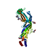
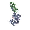
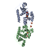
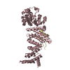
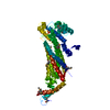
- PDB-4z35: Crystal Structure of Human Lysophosphatidic Acid Receptor 1 in co... -
+ Open data

- Basic information
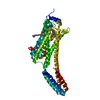
| Entry | Database: PDB / ID: 4z35 | ||||||
|---|---|---|---|---|---|---|---|
| Title | Crystal Structure of Human Lysophosphatidic Acid Receptor 1 in complex with ONO-9910539 | ||||||
 Components Components | Lysophosphatidic acid receptor 1,Soluble cytochrome b562 | ||||||
 Keywords Keywords | TRANSPORT PROTEIN/inhibitor / human lysophosphatidic acid receptor 1 (LPA1) / G-protein coupled receptor (GPCR) /  membrane protein / antagonist / endogenous ligand / PSI-biology / structural genomics / GPCR network / lipidic cubic phase (LCP) / compound design / polypharmacology / lipid receptor / TRANSPORT PROTEIN-inhibitor complex membrane protein / antagonist / endogenous ligand / PSI-biology / structural genomics / GPCR network / lipidic cubic phase (LCP) / compound design / polypharmacology / lipid receptor / TRANSPORT PROTEIN-inhibitor complex | ||||||
| Function / homology |  Function and homology information Function and homology informationcellular response to 1-oleoyl-sn-glycerol 3-phosphate / lysophosphatidic acid receptor activity / positive regulation of smooth muscle cell chemotaxis / calmodulin dependent kinase signaling pathway / Lysosphingolipid and LPA receptors / lysophosphatidic acid binding / negative regulation of cilium assembly / regulation of synaptic vesicle cycle / cellular response to oxygen levels / oligodendrocyte development ...cellular response to 1-oleoyl-sn-glycerol 3-phosphate / lysophosphatidic acid receptor activity / positive regulation of smooth muscle cell chemotaxis / calmodulin dependent kinase signaling pathway / Lysosphingolipid and LPA receptors / lysophosphatidic acid binding / negative regulation of cilium assembly / regulation of synaptic vesicle cycle / cellular response to oxygen levels / oligodendrocyte development / corpus callosum development / bleb assembly / regulation of metabolic process / optic nerve development / negative regulation of cAMP-mediated signaling / regulation of postsynaptic neurotransmitter receptor internalization / positive regulation of Rho protein signal transduction / positive regulation of dendritic spine development / : / G-protein alpha-subunit binding / GABA-ergic synapse / positive regulation of stress fiber assembly / myelination / adenylate cyclase-inhibiting G protein-coupled receptor signaling pathway / neurogenesis / cerebellum development / cell chemotaxis / electron transport chain / dendritic shaft / G protein-coupled receptor activity / PDZ domain binding / adenylate cyclase-activating G protein-coupled receptor signaling pathway / negative regulation of neuron projection development / presynaptic membrane / phospholipase C-activating G protein-coupled receptor signaling pathway / regulation of cell shape / positive regulation of cytosolic calcium ion concentration / G alpha (i) signalling events / G alpha (q) signalling events / postsynaptic membrane / positive regulation of canonical NF-kappaB signal transduction / dendritic spine / positive regulation of MAPK cascade / electron transfer activity / periplasmic space / endosome / iron ion binding / positive regulation of apoptotic process / G protein-coupled receptor signaling pathway / neuronal cell body / glutamatergic synapse / heme binding / cell surface / plasma membrane / cytoplasmSimilarity search - Function | ||||||
| Biological species |  Homo sapiens (human) Homo sapiens (human) Escherichia coli (E. coli) Escherichia coli (E. coli) | ||||||
| Method | X-RAY DIFFRACTION / SYNCHROTRON / MOLECULAR REPLACEMENT / Resolution: 2.9 Å | ||||||
 Authors Authors | Chrencik, J.E. / Roth, C.B. / Terakado, M. / Kurata, H. / Omi, R. / Kihara, Y. / Warshaviak, D. / Nakade, S. / Asmar-Rovira, G. / Mileni, M. ...Chrencik, J.E. / Roth, C.B. / Terakado, M. / Kurata, H. / Omi, R. / Kihara, Y. / Warshaviak, D. / Nakade, S. / Asmar-Rovira, G. / Mileni, M. / Mizuno, H. / Griffith, M.T. / Rodgers, C. / Han, G.W. / Velasquez, J. / Chun, J. / Stevens, R.C. / Hanson, M.A. / GPCR Network (GPCR) | ||||||
 Citation Citation | Journal: Cell / Year: 2015 Title: Crystal Structure of Antagonist Bound Human Lysophosphatidic Acid Receptor 1. Authors: Chrencik, J.E. / Roth, C.B. / Terakado, M. / Kurata, H. / Omi, R. / Kihara, Y. / Warshaviak, D. / Nakade, S. / Asmar-Rovira, G. / Mileni, M. / Mizuno, H. / Griffith, M.T. / Rodgers, C. / ...Authors: Chrencik, J.E. / Roth, C.B. / Terakado, M. / Kurata, H. / Omi, R. / Kihara, Y. / Warshaviak, D. / Nakade, S. / Asmar-Rovira, G. / Mileni, M. / Mizuno, H. / Griffith, M.T. / Rodgers, C. / Han, G.W. / Velasquez, J. / Chun, J. / Stevens, R.C. / Hanson, M.A. | ||||||
| History |
|
- Structure visualization
| Structure viewer | Molecule: MolmilJmol/JSmol |
|---|
- Downloads & links
-Download
| PDBx/mmCIF format | 4z35.cif.gz | 172 KB | Display | PDBx/mmCIF format |
|---|---|---|---|---|
| PDB format | pdb4z35.ent.gz | 133.3 KB | Display | PDB format |
| PDBx/mmJSON format | 4z35.json.gz | Tree view | PDBx/mmJSON format | |
| Others |  Other downloads Other downloads |
-Validation report
| Arichive directory | https://data.pdbj.org/pub/pdb/validation_reports/z3/4z35ftp://data.pdbj.org/pub/pdb/validation_reports/z3/4z35 | HTTPS FTP |
|---|
-Related structure data
| Related structure data |  4z34C 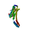 4z36C 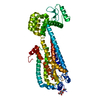 3v2yS 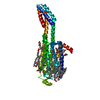 4eiyS C: citing same article ( S: Starting model for refinement |
|---|---|
| Similar structure data | |
| Other databases |


-Links
 PDBj
PDBj


- Assembly
| Deposited unit | 
| ||||||||
|---|---|---|---|---|---|---|---|---|---|
| 1 |
| ||||||||
| Unit cell |
| ||||||||
| Details | authors have indicated that the biological unit is unknown |
-Components
| #1: Protein | Mass: 52614.219 Da / Num. of mol.: 1 / Mutation: M1007W, H1102I, R1106L Source method: isolated from a genetically manipulated source Source: (gene. exp.) Homo sapiens (human), (gene. exp.) Escherichia coli (E. coli)Gene: LPAR1, EDG2, LPA1, cybC / Plasmid: pFASTBAC / Cell line (production host): sf9 / Production host:   Spodoptera frugiperda (fall armyworm) / References: UniProt: Q92633, UniProt: P0ABE7 Spodoptera frugiperda (fall armyworm) / References: UniProt: Q92633, UniProt: P0ABE7 |
|---|---|
| #2: Chemical | ChemComp-ON9 /   Mass: 563.638 Da / Num. of mol.: 1 / Source method: obtained synthetically / Formula: C32H37NO8 Mass: 563.638 Da / Num. of mol.: 1 / Source method: obtained synthetically / Formula: C32H37NO8 |
| #3: Chemical | ChemComp-1WV / (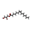  Mass: 300.434 Da / Num. of mol.: 1 / Source method: obtained synthetically / Formula: C17H32O4 Mass: 300.434 Da / Num. of mol.: 1 / Source method: obtained synthetically / Formula: C17H32O4 |
| #4: Water | ChemComp-HOH / Water Mass: 18.015 Da / Num. of mol.: 3 / Source method: isolated from a natural source / Formula: H2O Mass: 18.015 Da / Num. of mol.: 3 / Source method: isolated from a natural source / Formula: H2O |
-Experimental details
-Experiment
| Experiment | Method: X-RAY DIFFRACTION |
|---|
- Sample preparation
| Crystal | Density Matthews: 2.95 Å3/Da / Density % sol: 58.33 % |
|---|---|
| Crystal grow | Temperature: 293 K / Method: lipidic cubic phase / pH: 5.5 Details: 0.1 M sodium citrate (pH 5.5), 34 - 38% (v/v) PEG400 and 200 mM ammonium acetate |
-Data collection
| Diffraction |
| ||||||||||||||||||
|---|---|---|---|---|---|---|---|---|---|---|---|---|---|---|---|---|---|---|---|
| Diffraction source |
| ||||||||||||||||||
| Detector |
| ||||||||||||||||||
| Radiation |
| ||||||||||||||||||
| Radiation wavelength |
| ||||||||||||||||||
| Reflection | Resolution: 2.9→47 Å / Num. obs: 11627 / % possible obs: 78 % / Redundancy: 2.6 % / Biso Wilson estimate: 61.28 Å2 / Rmerge(I) obs: 0.14 / Net I/σ(I): 4 | ||||||||||||||||||
| Reflection shell | Resolution: 2.9→3.1 Å / Redundancy: 1.7 % / Rmerge(I) obs: 0.49 / Mean I/σ(I) obs: 1 / % possible all: 62 |

- Processing
| Software |
| ||||||||||||||||||||||||||||||||||||||||||||||||||||||||||||||||||||||||||||||||||||||||||||||||||||||||||||||||||
|---|---|---|---|---|---|---|---|---|---|---|---|---|---|---|---|---|---|---|---|---|---|---|---|---|---|---|---|---|---|---|---|---|---|---|---|---|---|---|---|---|---|---|---|---|---|---|---|---|---|---|---|---|---|---|---|---|---|---|---|---|---|---|---|---|---|---|---|---|---|---|---|---|---|---|---|---|---|---|---|---|---|---|---|---|---|---|---|---|---|---|---|---|---|---|---|---|---|---|---|---|---|---|---|---|---|---|---|---|---|---|---|---|---|---|---|
| Refinement | Method to determine structure: MOLECULAR REPLACEMENT Starting model: PDB ids 3V2Y and 4EIY Resolution: 2.9→30 Å / Cor.coef. Fo:Fc: 0.8895 / Cor.coef. Fo:Fc free: 0.8627 / Cross valid method: THROUGHOUT / σ(F): 0 / SU Rfree Blow DPI: 0.438
| ||||||||||||||||||||||||||||||||||||||||||||||||||||||||||||||||||||||||||||||||||||||||||||||||||||||||||||||||||
| Displacement parameters | Biso mean: 99.28 Å2
| ||||||||||||||||||||||||||||||||||||||||||||||||||||||||||||||||||||||||||||||||||||||||||||||||||||||||||||||||||
| Refine analyze | Luzzati coordinate error obs: 0.708 Å | ||||||||||||||||||||||||||||||||||||||||||||||||||||||||||||||||||||||||||||||||||||||||||||||||||||||||||||||||||
| Refinement step | Cycle: 1 / Resolution: 2.9→30 Å
| ||||||||||||||||||||||||||||||||||||||||||||||||||||||||||||||||||||||||||||||||||||||||||||||||||||||||||||||||||
| Refine LS restraints |
| ||||||||||||||||||||||||||||||||||||||||||||||||||||||||||||||||||||||||||||||||||||||||||||||||||||||||||||||||||
| LS refinement shell | Resolution: 2.9→3.18 Å / Total num. of bins used: 6
| ||||||||||||||||||||||||||||||||||||||||||||||||||||||||||||||||||||||||||||||||||||||||||||||||||||||||||||||||||
| Refinement TLS params. | Method: refined / Origin x: -0.3927 Å / Origin y: -18.87 Å / Origin z: 31.1438 Å
| ||||||||||||||||||||||||||||||||||||||||||||||||||||||||||||||||||||||||||||||||||||||||||||||||||||||||||||||||||
| Refinement TLS group | Selection details: { A|* } |
