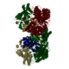
Entry Database : PDB / ID : 4yswTitle Structure of rat xanthine oxidoreductase, C-terminal deletion protein variant, NADH bound form Xanthine dehydrogenase/oxidase Keywords / / / / Function / homology Function Domain/homology Component
/ / / / / / / / / / / / / / / / / / / / / / / / / / / / / / / / / / / / / / / / / / / / / / / / / / / / / / / / / / / / / / / / / / / / / / / / / / / / / / / / / / / / / / / / / / / / / / / / / / / / / / / / / / / / / / / / / / / / / / / / / / / / / / / / / Biological species Rattus norvegicus (Norway rat)Method / / Resolution : 1.99 Å Authors Nishino, T. / Okamoto, K. / Kawaguchi, Y. / Matsumura, T. / Eger, B.T. / Pai, E.F. Journal : Febs J. / Year : 2015Title : The C-terminal peptide plays a role in the formation of an intermediate form during the transition between xanthine dehydrogenase and xanthine oxidase.Authors : Nishino, T. / Okamoto, K. / Kawaguchi, Y. / Matsumura, T. / Eger, B.T. / Pai, E.F. / Nishino, T. History Deposition Mar 17, 2015 Deposition site / Processing site Revision 1.0 Apr 22, 2015 Provider / Type Revision 1.1 Sep 9, 2015 Group Revision 1.2 Feb 5, 2020 Group / Derived calculations / Category / pdbx_struct_oper_listItem / _pdbx_struct_oper_list.symmetry_operationRevision 1.3 Mar 20, 2024 Group / Database references / Category / chem_comp_bond / database_2Item / _database_2.pdbx_database_accession
Show all Show less
 Movie
Movie Controller

 Yorodumi
 Open data
 Basic information
 Components
 Keywords
 OXIDOREDUCTASE /
  Function and homology information
 Authors
 Citation
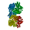
 Structure visualization
 Downloads & links
 Other downloads


 PDBj
PDBj


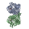
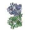
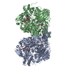
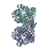
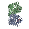
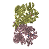
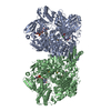
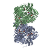
 Assembly
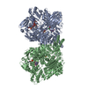


 Mass: 175.820 Da / Num. of mol.: 4 / Source method: obtained synthetically / Formula: Fe2S2
 Mass: 40.078 Da / Num. of mol.: 2 / Source method: obtained synthetically / Formula: Ca
 Mass: 785.550 Da / Num. of mol.: 2 / Source method: obtained synthetically / Formula: C27H33N9O15P2 / Comment: FAD*YM
 Mass: 665.441 Da / Num. of mol.: 2 / Source method: obtained synthetically / Formula: C21H29N7O14P2
 Mass: 61.017 Da / Num. of mol.: 2 / Source method: obtained synthetically / Formula: CHO3 / Comment: pH buffer*YM
 Mass: 168.110 Da / Num. of mol.: 2 / Source method: obtained synthetically / Formula: C5H4N4O3
 Mass: 92.094 Da / Num. of mol.: 2 / Source method: obtained synthetically / Formula: C3H8O3
 Sample preparation
 / Beamline: AR-NW12A / Wavelength: 1 Å
 Processing