 Movie
Movie Controller


+ Open data

- Basic information
| Entry | Database: PDB / ID: 4ysm | ||||||
|---|---|---|---|---|---|---|---|
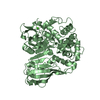
| Title | Calcium-Dependent Protein Kinase from Eimeria tenella | ||||||
 Components Components | Calmodulin-like domain protein kinase | ||||||
 Keywords Keywords |  TRANSFERASE / serine/threonine protein kinase / calcium-binding / ATP-binding TRANSFERASE / serine/threonine protein kinase / calcium-binding / ATP-binding | ||||||
| Function / homology |  Function and homology informationphosphorylation / protein serine/threonine kinase activity / calcium ion binding / ATP binding / membrane / cytoplasm Function and homology informationphosphorylation / protein serine/threonine kinase activity / calcium ion binding / ATP binding / membrane / cytoplasmSimilarity search - Function | ||||||
| Biological species |  Eimeria tenella (eukaryote) Eimeria tenella (eukaryote) | ||||||
| Method | X-RAY DIFFRACTION / SYNCHROTRON / MOLECULAR REPLACEMENT / molecular replacement / Resolution: 3.19 Å | ||||||
 Authors Authors | Merritt, E.A. | ||||||
| Funding support |  United States, 1items United States, 1items
| ||||||
 Citation Citation | Journal: to be published Title: CDPK is a druggable target in the apicomplexan parasite Eimeria Authors: Ojo, K.K. / Vidadala, R. / Maly, D.J. / Van Voorhis, W.C. / Merritt, E.A. | ||||||
| History |
|
- Structure visualization
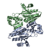
| Structure viewer | Molecule: MolmilJmol/JSmol |
|---|
- Downloads & links
-Download
| PDBx/mmCIF format | 4ysm.cif.gz | 392.1 KB | Display | PDBx/mmCIF format |
|---|---|---|---|---|
| PDB format | pdb4ysm.ent.gz | 322.4 KB | Display | PDB format |
| PDBx/mmJSON format | 4ysm.json.gz | Tree view | PDBx/mmJSON format | |
| Others |  Other downloads Other downloads |
-Validation report
| Arichive directory | https://data.pdbj.org/pub/pdb/validation_reports/ys/4ysmftp://data.pdbj.org/pub/pdb/validation_reports/ys/4ysm | HTTPS FTP |
|---|
-Related structure data
| Related structure data |  4ysjC 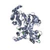 4yuqC 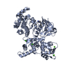 4yzbC 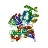 3q5iS C: citing same article ( S: Starting model for refinement |
|---|---|
| Similar structure data |


-Links
 PDBj
PDBj- Assembly
| Deposited unit | 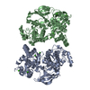
| ||||||||||||||||||
|---|---|---|---|---|---|---|---|---|---|---|---|---|---|---|---|---|---|---|---|
| 1 | 
| ||||||||||||||||||
| 2 | 
| ||||||||||||||||||
| Unit cell |
| ||||||||||||||||||
| Noncrystallographic symmetry (NCS) | NCS domain:
NCS domain segments: Component-ID: 0 / Ens-ID: 1 / Beg auth comp-ID: ASP / Beg label comp-ID: ASP / End auth comp-ID: LEU / End label comp-ID: LEU / Refine code: 0 / Auth seq-ID: 7 - 481 / Label seq-ID: 11 - 485
|
-Components
| #1: Protein | Mass: 55668.504 Da / Num. of mol.: 2 / Mutation: A410D Source method: isolated from a genetically manipulated source Details: cleaved N-terminal His-tag / Source: (gene. exp.) Eimeria tenella (eukaryote) / Plasmid: AVA0421 / Production host:  Escherichia coli (E. coli) / Strain (production host): BL21(DE3) / References: UniProt: Q3HNM4 Escherichia coli (E. coli) / Strain (production host): BL21(DE3) / References: UniProt: Q3HNM4#2: Chemical | ChemComp-CA /   Mass: 40.078 Da / Num. of mol.: 8 / Source method: obtained synthetically / Formula: Ca Mass: 40.078 Da / Num. of mol.: 8 / Source method: obtained synthetically / Formula: Ca#3: Water | ChemComp-HOH / | Water Mass: 18.015 Da / Num. of mol.: 2 / Source method: isolated from a natural source / Formula: H2O Mass: 18.015 Da / Num. of mol.: 2 / Source method: isolated from a natural source / Formula: H2OSequence details | AUTHOR STATED THAT THE UNP REFERENCE Q3HNM4 IS INCORRECT AT RESIDUE 410. THE RESIDUE IDENTITY OF ...AUTHOR STATED THAT THE UNP REFERENCE Q3HNM4 IS INCORRECT AT RESIDUE 410. THE RESIDUE IDENTITY OF 410 SHOULD BE ASP INSTEAD OF ALA BECAUSE THIS RESIDUE IS IN BINDING TO CALCIUM AND CONSERVED ACROSS THE LARGER FAMILY OF CDPKS. | |
|---|
-Experimental details
-Experiment
| Experiment | Method: X-RAY DIFFRACTION / Number of used crystals: 1 |
|---|
- Sample preparation
| Crystal | Density Matthews: 2.55 Å3/Da / Density % sol: 51.72 % |
|---|---|
| Crystal grow | Temperature: 298 K / Method: vapor diffusion, hanging drop Details: protein buffer: 25 mM HEPES pH 7.0, 5% glycerol, 500 mM NaCl, 2 mM DTT, 175 uM EtCDPK1; crystallization buffer: 100 mM BIS/TRIS pH 6.5, 23% PEG 3350 |
-Data collection
| Diffraction | Mean temperature: 100 K | |||||||||||||||||||||||||||
|---|---|---|---|---|---|---|---|---|---|---|---|---|---|---|---|---|---|---|---|---|---|---|---|---|---|---|---|---|
| Diffraction source | Source: SYNCHROTRON / Site: SSRL / Beamline: BL11-1 / Wavelength: 0.97945 Å | |||||||||||||||||||||||||||
| Detector | Type: PSI PILATUS 6M / Detector: PIXEL / Date: Nov 25, 2014 | |||||||||||||||||||||||||||
| Radiation | Protocol: SINGLE WAVELENGTH / Monochromatic (M) / Laue (L): M / Scattering type: x-ray | |||||||||||||||||||||||||||
| Radiation wavelength | Wavelength: 0.97945 Å / Relative weight: 1 | |||||||||||||||||||||||||||
| Reflection | Resolution: 3.19→38.84 Å / Num. obs: 18399 / % possible obs: 98.8 % / Redundancy: 6.9 % / Biso Wilson estimate: 78 Å2 / CC1/2: 0.976 / Rmerge(I) obs: 0.497 / Rpim(I) all: 0.203 / Net I/σ(I): 5.6 / Num. measured all: 127487 / Scaling rejects: 2 | |||||||||||||||||||||||||||
| Reflection shell | Diffraction-ID: 1 / Rejects: 0
|
-Phasing
| Phasing | Method: molecular replacement |
|---|
- Processing
| Software |
| |||||||||||||||||||||||||||||||||||||||||||||||||||||||||||||||||||||||||||||||||||||||||||||||||||||||||||||||||||||||||||||||||||||||||||||||||||||||||||||||||||||||||||||||||||||||||||||||||||||||||||||||||||||||||||||||||||||||||||||||||||||||||||||||||||||||||||||||||||||||||||||||||||||||||||||||||||||||||||||||||||||
|---|---|---|---|---|---|---|---|---|---|---|---|---|---|---|---|---|---|---|---|---|---|---|---|---|---|---|---|---|---|---|---|---|---|---|---|---|---|---|---|---|---|---|---|---|---|---|---|---|---|---|---|---|---|---|---|---|---|---|---|---|---|---|---|---|---|---|---|---|---|---|---|---|---|---|---|---|---|---|---|---|---|---|---|---|---|---|---|---|---|---|---|---|---|---|---|---|---|---|---|---|---|---|---|---|---|---|---|---|---|---|---|---|---|---|---|---|---|---|---|---|---|---|---|---|---|---|---|---|---|---|---|---|---|---|---|---|---|---|---|---|---|---|---|---|---|---|---|---|---|---|---|---|---|---|---|---|---|---|---|---|---|---|---|---|---|---|---|---|---|---|---|---|---|---|---|---|---|---|---|---|---|---|---|---|---|---|---|---|---|---|---|---|---|---|---|---|---|---|---|---|---|---|---|---|---|---|---|---|---|---|---|---|---|---|---|---|---|---|---|---|---|---|---|---|---|---|---|---|---|---|---|---|---|---|---|---|---|---|---|---|---|---|---|---|---|---|---|---|---|---|---|---|---|---|---|---|---|---|---|---|---|---|---|---|---|---|---|---|---|---|---|---|---|---|---|---|---|---|---|---|---|---|---|---|---|---|---|---|---|---|---|---|---|---|---|---|---|---|---|---|---|---|---|---|---|---|---|---|---|---|---|---|---|---|---|---|---|---|---|---|---|---|---|---|---|---|
| Refinement | Method to determine structure: MOLECULAR REPLACEMENT Starting model: PDB entry 3q5i Resolution: 3.19→38.84 Å / Cor.coef. Fo:Fc: 0.915 / Cor.coef. Fo:Fc free: 0.86 / WRfactor Rfree: 0.2554 / WRfactor Rwork: 0.2084 / FOM work R set: 0.7306 / SU B: 81.955 / SU ML: 0.614 / SU R Cruickshank DPI: 0.496 / SU Rfree: 0.6056 / Cross valid method: THROUGHOUT / σ(F): 0 / ESU R Free: 0.606 / Stereochemistry target values: MAXIMUM LIKELIHOOD Details: HYDROGENS HAVE BEEN ADDED IN THE RIDING POSITIONS U VALUES : WITH TLS ADDED
| |||||||||||||||||||||||||||||||||||||||||||||||||||||||||||||||||||||||||||||||||||||||||||||||||||||||||||||||||||||||||||||||||||||||||||||||||||||||||||||||||||||||||||||||||||||||||||||||||||||||||||||||||||||||||||||||||||||||||||||||||||||||||||||||||||||||||||||||||||||||||||||||||||||||||||||||||||||||||||||||||||||
| Solvent computation | Ion probe radii: 0.8 Å / Shrinkage radii: 0.8 Å / VDW probe radii: 1.2 Å / Solvent model: MASK | |||||||||||||||||||||||||||||||||||||||||||||||||||||||||||||||||||||||||||||||||||||||||||||||||||||||||||||||||||||||||||||||||||||||||||||||||||||||||||||||||||||||||||||||||||||||||||||||||||||||||||||||||||||||||||||||||||||||||||||||||||||||||||||||||||||||||||||||||||||||||||||||||||||||||||||||||||||||||||||||||||||
| Displacement parameters | Biso max: 163.04 Å2 / Biso mean: 73.509 Å2 / Biso min: 26.82 Å2
| |||||||||||||||||||||||||||||||||||||||||||||||||||||||||||||||||||||||||||||||||||||||||||||||||||||||||||||||||||||||||||||||||||||||||||||||||||||||||||||||||||||||||||||||||||||||||||||||||||||||||||||||||||||||||||||||||||||||||||||||||||||||||||||||||||||||||||||||||||||||||||||||||||||||||||||||||||||||||||||||||||||
| Refinement step | Cycle: final / Resolution: 3.19→38.84 Å
| |||||||||||||||||||||||||||||||||||||||||||||||||||||||||||||||||||||||||||||||||||||||||||||||||||||||||||||||||||||||||||||||||||||||||||||||||||||||||||||||||||||||||||||||||||||||||||||||||||||||||||||||||||||||||||||||||||||||||||||||||||||||||||||||||||||||||||||||||||||||||||||||||||||||||||||||||||||||||||||||||||||
| Refine LS restraints |
| |||||||||||||||||||||||||||||||||||||||||||||||||||||||||||||||||||||||||||||||||||||||||||||||||||||||||||||||||||||||||||||||||||||||||||||||||||||||||||||||||||||||||||||||||||||||||||||||||||||||||||||||||||||||||||||||||||||||||||||||||||||||||||||||||||||||||||||||||||||||||||||||||||||||||||||||||||||||||||||||||||||
| Refine LS restraints NCS | Ens-ID: 1 / Number: 29127 / Refine-ID: X-RAY DIFFRACTION / Type: interatomic distance / Rms dev position: 0.11 Å / Weight position: 0.05
| |||||||||||||||||||||||||||||||||||||||||||||||||||||||||||||||||||||||||||||||||||||||||||||||||||||||||||||||||||||||||||||||||||||||||||||||||||||||||||||||||||||||||||||||||||||||||||||||||||||||||||||||||||||||||||||||||||||||||||||||||||||||||||||||||||||||||||||||||||||||||||||||||||||||||||||||||||||||||||||||||||||
| LS refinement shell | Resolution: 3.194→3.277 Å / Total num. of bins used: 20
| |||||||||||||||||||||||||||||||||||||||||||||||||||||||||||||||||||||||||||||||||||||||||||||||||||||||||||||||||||||||||||||||||||||||||||||||||||||||||||||||||||||||||||||||||||||||||||||||||||||||||||||||||||||||||||||||||||||||||||||||||||||||||||||||||||||||||||||||||||||||||||||||||||||||||||||||||||||||||||||||||||||
| Refinement TLS params. | Method: refined / Refine-ID: X-RAY DIFFRACTION
| |||||||||||||||||||||||||||||||||||||||||||||||||||||||||||||||||||||||||||||||||||||||||||||||||||||||||||||||||||||||||||||||||||||||||||||||||||||||||||||||||||||||||||||||||||||||||||||||||||||||||||||||||||||||||||||||||||||||||||||||||||||||||||||||||||||||||||||||||||||||||||||||||||||||||||||||||||||||||||||||||||||
| Refinement TLS group |
|
