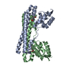
symbiont-mediated suppression of host JAK-STAT cascade via inhibition of host IRF9 activity / symbiont-mediated activation of of host transcription / : / regulation of lipid kinase activity / : / Transcription of E2F targets under negative control by p107 (RBL1) and p130 (RBL2) in complex with HDAC1 / Transcription of E2F targets under negative control by DREAM complex / negative regulation of G1/S transition of mitotic cell cycle / positive regulation of actin filament polymerization / G1/S-Specific Transcription ...symbiont-mediated suppression of host JAK-STAT cascade via inhibition of host IRF9 activity / symbiont-mediated activation of of host transcription / : / regulation of lipid kinase activity / : / Transcription of E2F targets under negative control by p107 (RBL1) and p130 (RBL2) in complex with HDAC1 / Transcription of E2F targets under negative control by DREAM complex / negative regulation of G1/S transition of mitotic cell cycle / positive regulation of actin filament polymerization / G1/S-Specific Transcription / cadmium ion binding / symbiont-mediated perturbation of host cell cycle G1/S transition checkpoint / negative regulation of cellular senescence / G0 and Early G1 / viral process / TP53 Regulates Transcription of Genes Involved in G2 Cell Cycle Arrest / promoter-specific chromatin binding / RNA polymerase II transcription regulatory region sequence-specific DNA binding / SMAD2/SMAD3:SMAD4 heterotrimer regulates transcription / Cyclin D associated events in G1 / chromatin organization / DNA-binding transcription factor binding / host cell cytoplasm / transcription regulator complex / cell differentiation / cell cycle / DNA-binding transcription factor activity / protein domain specific binding / negative regulation of gene expression / DNA-templated transcription / symbiont-mediated suppression of host type I interferon-mediated signaling pathway / host cell nucleus / chromatin / negative regulation of transcription by RNA polymerase II / DNA binding / zinc ion binding / nucleoplasm Similarity search - Function
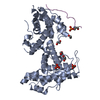
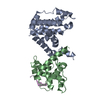
Papillomavirus E7 / E7 protein, Early protein / Rb C-terminal domain / Retinoblastoma-associated protein, B-box / Retinoblastoma-associated protein, A-box / Retinoblastoma-associated protein, C-terminal / Retinoblastoma-associated protein, N-terminal / Retinoblastoma protein family / Retinoblastoma-associated protein B domain / Retinoblastoma-associated protein A domain ...Papillomavirus E7 / E7 protein, Early protein / Rb C-terminal domain / Retinoblastoma-associated protein, B-box / Retinoblastoma-associated protein, A-box / Retinoblastoma-associated protein, C-terminal / Retinoblastoma-associated protein, N-terminal / Retinoblastoma protein family / Retinoblastoma-associated protein B domain / Retinoblastoma-associated protein A domain / Domain of unknown function (DUF3452) / Domain of unknown function (DUF3452) / Retinoblastoma-associated protein A domain / Rb C-terminal domain / Cyclin-like / Cyclin A; domain 1 / Cyclin-like / domain present in cyclins, TFIIB and Retinoblastoma / Cyclin-like superfamily / Orthogonal Bundle / Mainly Alpha Similarity search - Domain/homology
In the structure databanks used in Yorodumi, some data are registered as the other names, "COVID-19 virus" and "2019-nCoV". Here are the details of the virus and the list of structure data.
Jan 31, 2019. EMDB accession codes are about to change! (news from PDBe EMDB page)
EMDB accession codes are about to change! (news from PDBe EMDB page)
The allocation of 4 digits for EMDB accession codes will soon come to an end. Whilst these codes will remain in use, new EMDB accession codes will include an additional digit and will expand incrementally as the available range of codes is exhausted. The current 4-digit format prefixed with “EMD-” (i.e. EMD-XXXX) will advance to a 5-digit format (i.e. EMD-XXXXX), and so on. It is currently estimated that the 4-digit codes will be depleted around Spring 2019, at which point the 5-digit format will come into force.
The EM Navigator/Yorodumi systems omit the EMD- prefix.
Related info.:Q: What is EMD? / ID/Accession-code notation in Yorodumi/EM Navigator
Yorodumi is a browser for structure data from EMDB, PDB, SASBDB, etc.
This page is also the successor to EM Navigator detail page, and also detail information page/front-end page for Omokage search.
The word "yorodu" (or yorozu) is an old Japanese word meaning "ten thousand". "mi" (miru) is to see.
Related info.:EMDB / PDB / SASBDB / Comparison of 3 databanks / Yorodumi Search / Aug 31, 2016. New EM Navigator & Yorodumi / Yorodumi Papers / Jmol/JSmol / Function and homology information / Changes in new EM Navigator and Yorodumi
 Movie
Movie Controller


 Open data
 Basic information
 Components
 Keywords
 TRANSCRIPTION / Pocket protein / Cyclin Box /
  Function and homology information

 Authors
 United States, 1items
  Citation
 Structure visualization
 Downloads & links
 Other downloads
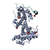


 PDBj
PDBj


 Assembly
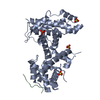


 Mass: 96.063 Da / Num. of mol.: 4 / Source method: obtained synthetically / Formula: SO4
 Mass: 18.015 Da / Num. of mol.: 111 / Source method: isolated from a natural source / Formula: H2O
 Sample preparation
 Processing