 Movie
Movie Controller


+ Open data

- Basic information
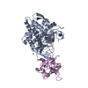
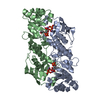
| Entry | Database: PDB / ID: 4yom | |||||||||||||||
|---|---|---|---|---|---|---|---|---|---|---|---|---|---|---|---|---|
| Title | Structure of SAD kinase | |||||||||||||||
 Components Components | (Serine/threonine-protein kinase BRSK2) x 2 | |||||||||||||||
 Keywords Keywords |  TRANSFERASE / kinase domain / UBA domain / KA1 domain TRANSFERASE / kinase domain / UBA domain / KA1 domain | |||||||||||||||
| Function / homology |  Function and homology information Function and homology informationdistal axon / microtubule cytoskeleton organization involved in establishment of planar polarity / : / regulation of insulin secretion involved in cellular response to glucose stimulus / tau-protein kinase / regulation of neuron projection development / regulation of axonogenesis / establishment of cell polarity / tau-protein kinase activity / exocytosis ...distal axon / microtubule cytoskeleton organization involved in establishment of planar polarity / : / regulation of insulin secretion involved in cellular response to glucose stimulus / tau-protein kinase / regulation of neuron projection development / regulation of axonogenesis / establishment of cell polarity / tau-protein kinase activity / exocytosis / ERAD pathway / intrinsic apoptotic signaling pathway in response to endoplasmic reticulum stress / axonogenesis / neuron projection morphogenesis / neuron differentiation / G2/M transition of mitotic cell cycle / ATPase binding / peptidyl-serine phosphorylation / non-specific serine/threonine protein kinase / intracellular signal transduction / cell division / protein phosphorylation / protein serine kinase activity / protein serine/threonine kinase activity / centrosome / protein kinase binding / perinuclear region of cytoplasm / magnesium ion binding / endoplasmic reticulum / ATP binding / cytoplasmSimilarity search - Function | |||||||||||||||
| Biological species |  Mus musculus (house mouse) Mus musculus (house mouse) | |||||||||||||||
| Method | X-RAY DIFFRACTION / SYNCHROTRON / MOLECULAR REPLACEMENT / Resolution: 2.49 Å | |||||||||||||||
 Authors Authors | Wu, J.X. / Wang, J. / Chen, L. / Wang, Z.X. / Wu, J.W. | |||||||||||||||
| Funding support |  China, 4items China, 4items
| |||||||||||||||
 Citation Citation | Journal: Nat Commun / Year: 2015 Title: Structural insight into the mechanism of synergistic autoinhibition of SAD kinases Authors: Wu, J.X. / Cheng, Y.S. / Wang, J. / Chen, L. / Ding, M. / Wu, J.W. | |||||||||||||||
| History |
|
- Structure visualization
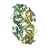
| Structure viewer | Molecule: MolmilJmol/JSmol |
|---|
- Downloads & links
-Download
| PDBx/mmCIF format | 4yom.cif.gz | 190.8 KB | Display | PDBx/mmCIF format |
|---|---|---|---|---|
| PDB format | pdb4yom.ent.gz | 150.3 KB | Display | PDB format |
| PDBx/mmJSON format | 4yom.json.gz | Tree view | PDBx/mmJSON format | |
| Others |  Other downloads Other downloads |
-Validation report
| Arichive directory | https://data.pdbj.org/pub/pdb/validation_reports/yo/4yomftp://data.pdbj.org/pub/pdb/validation_reports/yo/4yom | HTTPS FTP |
|---|
-Related structure data
| Related structure data | 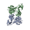 4ynzC  3oseS C: citing same article ( S: Starting model for refinement |
|---|---|
| Similar structure data |


-Links
 PDBj
PDBj- Assembly
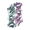
| Deposited unit | 
| ||||||||
|---|---|---|---|---|---|---|---|---|---|
| 1 |
| ||||||||
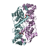
| Unit cell |
|
-Components
| #1: Protein | Mass: 40171.516 Da / Num. of mol.: 1 / Fragment: UNP residues 1-342 Source method: isolated from a genetically manipulated source Source: (gene. exp.) Mus musculus (house mouse) / Gene: Brsk2 / Plasmid: PET21b / Production host:  Escherichia coli BL21(DE3) (bacteria) / Strain (production host): BL21(DE3) / References: UniProt: Q69Z98, tau-protein kinase Escherichia coli BL21(DE3) (bacteria) / Strain (production host): BL21(DE3) / References: UniProt: Q69Z98, tau-protein kinase | ||||
|---|---|---|---|---|---|
| #2: Protein | Mass: 16215.451 Da / Num. of mol.: 1 / Fragment: UNP residues 519-653 Source method: isolated from a genetically manipulated source Source: (gene. exp.) Mus musculus (house mouse) / Gene: Brsk2 / Plasmid: PET21b / Production host: Escherichia coli BL21(DE3) (bacteria) / Strain (production host): BL21(DE3) / References: UniProt: Q69Z98, tau-protein kinase | ||||
| #3: Chemical | Ethylene glycol  Mass: 62.068 Da / Num. of mol.: 2 / Source method: obtained synthetically / Formula: C2H6O2 Mass: 62.068 Da / Num. of mol.: 2 / Source method: obtained synthetically / Formula: C2H6O2#4: Water | ChemComp-HOH / | Water Mass: 18.015 Da / Num. of mol.: 67 / Source method: isolated from a natural source / Formula: H2O Mass: 18.015 Da / Num. of mol.: 67 / Source method: isolated from a natural source / Formula: H2OSequence details | The sequence of chain A is Isoform 4 SADA-alpha. | |
-Experimental details
-Experiment
| Experiment | Method: X-RAY DIFFRACTION |
|---|
- Sample preparation
| Crystal | Density Matthews: 3.05 Å3/Da / Density % sol: 59.62 % |
|---|---|
| Crystal grow | Temperature: 277 K / Method: vapor diffusion, hanging drop / pH: 5.2 / Details: 0.1 M Na Citrate, 1.0 M Na Malonate |
-Data collection
| Diffraction | Mean temperature: 100 K |
|---|---|
| Diffraction source | Source: SYNCHROTRON / Site: SSRF / Beamline: BL17U / Wavelength: 0.9798 Å |
| Detector | Type: ADSC QUANTUM 315r / Detector: CCD / Date: Jun 5, 2012 |
| Radiation | Monochromator: double crystal / Protocol: SINGLE WAVELENGTH / Monochromatic (M) / Laue (L): M / Scattering type: x-ray |
| Radiation wavelength | Wavelength: 0.9798 Å / Relative weight: 1 |
| Reflection | Resolution: 2.49→50 Å / Num. obs: 23808 / % possible obs: 100 % / Redundancy: 7.5 % / Biso Wilson estimate: 56.79 Å2 / Rmerge(I) obs: 0.087 / Net I/σ(I): 26.9 |
| Reflection shell | Resolution: 2.49→2.58 Å / Redundancy: 7.5 % / Rmerge(I) obs: 0.719 / Mean I/σ(I) obs: 4.8 / % possible all: 100 |
- Processing
| Software |
| |||||||||||||||||||||||||||||||||||||||||||||||||||||||||||||||||||||||||||||||||||||||||||||||||||||||||||||||||||||||||||||||||||||||||||||||||||||||||||||||||||||||||||||||||||||||||||||||||||||||||||||||||||||||||||||||||
|---|---|---|---|---|---|---|---|---|---|---|---|---|---|---|---|---|---|---|---|---|---|---|---|---|---|---|---|---|---|---|---|---|---|---|---|---|---|---|---|---|---|---|---|---|---|---|---|---|---|---|---|---|---|---|---|---|---|---|---|---|---|---|---|---|---|---|---|---|---|---|---|---|---|---|---|---|---|---|---|---|---|---|---|---|---|---|---|---|---|---|---|---|---|---|---|---|---|---|---|---|---|---|---|---|---|---|---|---|---|---|---|---|---|---|---|---|---|---|---|---|---|---|---|---|---|---|---|---|---|---|---|---|---|---|---|---|---|---|---|---|---|---|---|---|---|---|---|---|---|---|---|---|---|---|---|---|---|---|---|---|---|---|---|---|---|---|---|---|---|---|---|---|---|---|---|---|---|---|---|---|---|---|---|---|---|---|---|---|---|---|---|---|---|---|---|---|---|---|---|---|---|---|---|---|---|---|---|---|---|---|---|---|---|---|---|---|---|---|---|---|---|---|---|---|---|---|
| Refinement | Method to determine structure: MOLECULAR REPLACEMENT Starting model: 3OSE Resolution: 2.49→34.579 Å / SU ML: 0.3 / Cross valid method: FREE R-VALUE / σ(F): 1.34 / Phase error: 25.75 / Stereochemistry target values: ML
| |||||||||||||||||||||||||||||||||||||||||||||||||||||||||||||||||||||||||||||||||||||||||||||||||||||||||||||||||||||||||||||||||||||||||||||||||||||||||||||||||||||||||||||||||||||||||||||||||||||||||||||||||||||||||||||||||
| Solvent computation | Shrinkage radii: 0.73 Å / VDW probe radii: 1 Å / Solvent model: FLAT BULK SOLVENT MODEL / Bsol: 35.315 Å2 / ksol: 0.341 e/Å3 | |||||||||||||||||||||||||||||||||||||||||||||||||||||||||||||||||||||||||||||||||||||||||||||||||||||||||||||||||||||||||||||||||||||||||||||||||||||||||||||||||||||||||||||||||||||||||||||||||||||||||||||||||||||||||||||||||
| Displacement parameters | Biso mean: 54.27 Å2
| |||||||||||||||||||||||||||||||||||||||||||||||||||||||||||||||||||||||||||||||||||||||||||||||||||||||||||||||||||||||||||||||||||||||||||||||||||||||||||||||||||||||||||||||||||||||||||||||||||||||||||||||||||||||||||||||||
| Refinement step | Cycle: LAST / Resolution: 2.49→34.579 Å
| |||||||||||||||||||||||||||||||||||||||||||||||||||||||||||||||||||||||||||||||||||||||||||||||||||||||||||||||||||||||||||||||||||||||||||||||||||||||||||||||||||||||||||||||||||||||||||||||||||||||||||||||||||||||||||||||||
| Refine LS restraints |
| |||||||||||||||||||||||||||||||||||||||||||||||||||||||||||||||||||||||||||||||||||||||||||||||||||||||||||||||||||||||||||||||||||||||||||||||||||||||||||||||||||||||||||||||||||||||||||||||||||||||||||||||||||||||||||||||||
| LS refinement shell |
| |||||||||||||||||||||||||||||||||||||||||||||||||||||||||||||||||||||||||||||||||||||||||||||||||||||||||||||||||||||||||||||||||||||||||||||||||||||||||||||||||||||||||||||||||||||||||||||||||||||||||||||||||||||||||||||||||
| Refinement TLS params. | Method: refined / Refine-ID: X-RAY DIFFRACTION
| |||||||||||||||||||||||||||||||||||||||||||||||||||||||||||||||||||||||||||||||||||||||||||||||||||||||||||||||||||||||||||||||||||||||||||||||||||||||||||||||||||||||||||||||||||||||||||||||||||||||||||||||||||||||||||||||||
| Refinement TLS group |
|
