 Movie
Movie Controller


+ Open data

- Basic information
| Entry | Database: PDB / ID: 4yni | ||||||
|---|---|---|---|---|---|---|---|
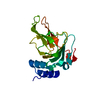
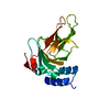
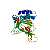
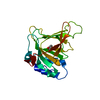
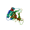
| Title | Iron free succinate bound rat cysteine dioxygenase | ||||||
 Components Components | Cysteine dioxygenase type 1 | ||||||
 Keywords Keywords | OXIDOREDUCTASE / non-heme mono-iron / Cupin / succinate / iron free | ||||||
| Function / homology |  Function and homology information Function and homology informationL-cysteine metabolic process / Degradation of cysteine and homocysteine / taurine biosynthetic process / cysteine dioxygenase / cysteine dioxygenase activity / L-cysteine catabolic process / cysteine metabolic process / response to glucagon / nickel cation binding / response to amino acid ...L-cysteine metabolic process / Degradation of cysteine and homocysteine / taurine biosynthetic process / cysteine dioxygenase / cysteine dioxygenase activity / L-cysteine catabolic process / cysteine metabolic process / response to glucagon / nickel cation binding / response to amino acid / response to glucocorticoid / response to cAMP / response to organonitrogen compound / lactation / ferrous iron binding / response to ethanol / zinc ion binding / cytosolSimilarity search - Function | ||||||
| Biological species |  Rattus norvegicus (Norway rat) Rattus norvegicus (Norway rat) | ||||||
| Method | X-RAY DIFFRACTION / MOLECULAR REPLACEMENT / molecular replacement / Resolution: 2.404 Å | ||||||
 Authors Authors | Fellner, M. / Tchesnokov, E.P. / Jameson, G.N.L. / Wilbanks, S.M. | ||||||
 Citation Citation | Journal: To be published Title: Iron free succinate bound rat cysteine dioxygenase Authors: Fellner, M. / Tchesnokov, E.P. / Jameson, G.N.L. / Wilbanks, S.M. | ||||||
| History |
|
- Structure visualization
| Structure viewer | Molecule: MolmilJmol/JSmol |
|---|
- Downloads & links
-Download
| PDBx/mmCIF format | 4yni.cif.gz | 56.2 KB | Display | PDBx/mmCIF format |
|---|---|---|---|---|
| PDB format | pdb4yni.ent.gz | 38.7 KB | Display | PDB format |
| PDBx/mmJSON format | 4yni.json.gz | Tree view | PDBx/mmJSON format | |
| Others |  Other downloads Other downloads |
-Validation report
| Arichive directory | https://data.pdbj.org/pub/pdb/validation_reports/yn/4yniftp://data.pdbj.org/pub/pdb/validation_reports/yn/4yni | HTTPS FTP |
|---|
-Related structure data
| Related structure data |  4kwjS S: Starting model for refinement |
|---|---|
| Similar structure data |


-Links
 PDBj
PDBj- Assembly
| Deposited unit | 
| ||||||||
|---|---|---|---|---|---|---|---|---|---|
| 1 |
| ||||||||
| Unit cell |
|
-Components
| #1: Protein | / Cysteine dioxygenase type I / CDO-I Mass: 24259.195 Da / Num. of mol.: 1 Source method: isolated from a genetically manipulated source Source: (gene. exp.) Rattus norvegicus (Norway rat) / Gene: Cdo1 / Plasmid: pPR-IBA1/RatCDO/WT / Production host:  Escherichia coli (E. coli) / Strain (production host): BL21(DE3)pLysS / References: UniProt: P21816, cysteine dioxygenase Escherichia coli (E. coli) / Strain (production host): BL21(DE3)pLysS / References: UniProt: P21816, cysteine dioxygenase |
|---|---|
| #2: Chemical | ChemComp-NA /   Mass: 22.990 Da / Num. of mol.: 1 / Source method: obtained synthetically / Formula: Na Mass: 22.990 Da / Num. of mol.: 1 / Source method: obtained synthetically / Formula: Na |
| #3: Chemical | ChemComp-SIN / Succinic acid  Mass: 118.088 Da / Num. of mol.: 1 / Source method: obtained synthetically / Formula: C4H6O4 Mass: 118.088 Da / Num. of mol.: 1 / Source method: obtained synthetically / Formula: C4H6O4 |
| #4: Water | ChemComp-HOH / Water Mass: 18.015 Da / Num. of mol.: 87 / Source method: isolated from a natural source / Formula: H2O Mass: 18.015 Da / Num. of mol.: 87 / Source method: isolated from a natural source / Formula: H2O |
-Experimental details
-Experiment
| Experiment | Method: X-RAY DIFFRACTION / Number of used crystals: 1 |
|---|
- Sample preparation
| Crystal | Density Matthews: 2.09 Å3/Da / Density % sol: 41.15 % |
|---|---|
| Crystal grow | Temperature: 291 K / Method: vapor diffusion, hanging drop / pH: 5.2 Details: Hanging drops of 1 microL of 15 mg/mL WT-CDO (10mM sodiumphosphate, 20mM NaCl pH 7.5) and 3 microL reservoir buffer were allowed to equilibrate above the reservoir buffer (25% (w/v) ...Details: Hanging drops of 1 microL of 15 mg/mL WT-CDO (10mM sodiumphosphate, 20mM NaCl pH 7.5) and 3 microL reservoir buffer were allowed to equilibrate above the reservoir buffer (25% (w/v) polyethylene glycol 1500, 13 mM succinate, 44 mM mono sodium phosphate, 44 mM glycine) and 0.6 microL WT-CDO seeds (grown in the reservoir buffer) |
-Data collection
| Diffraction | Mean temperature: 93 K | |||||||||||||||||||||||||||
|---|---|---|---|---|---|---|---|---|---|---|---|---|---|---|---|---|---|---|---|---|---|---|---|---|---|---|---|---|
| Diffraction source | Source: ROTATING ANODE / Type: RIGAKU / Wavelength: 1.54187 Å | |||||||||||||||||||||||||||
| Detector | Type: RIGAKU RAXIS IV++ / Detector: IMAGE PLATE / Date: Dec 2, 2013 / Details: Osmic VariMax optics | |||||||||||||||||||||||||||
| Radiation | Monochromator: Osmic VariMax optics / Protocol: SINGLE WAVELENGTH / Monochromatic (M) / Laue (L): M / Scattering type: x-ray | |||||||||||||||||||||||||||
| Radiation wavelength | Wavelength: 1.54187 Å / Relative weight: 1 | |||||||||||||||||||||||||||
| Reflection | Resolution: 2.4→41.89 Å / Num. obs: 15515 / % possible obs: 100 % / Redundancy: 10 % / Biso Wilson estimate: 36.89 Å2 / CC1/2: 0.996 / Rmerge(I) obs: 0.134 / Rpim(I) all: 0.044 / Net I/σ(I): 11.5 / Num. measured all: 85493 / Scaling rejects: 168 | |||||||||||||||||||||||||||
| Reflection shell | Diffraction-ID: 1 / Rejects: 0
|
-Phasing
| Phasing | Method: molecular replacement |
|---|
- Processing
| Software |
| |||||||||||||||||||||||||||||||||||||||||||||||||
|---|---|---|---|---|---|---|---|---|---|---|---|---|---|---|---|---|---|---|---|---|---|---|---|---|---|---|---|---|---|---|---|---|---|---|---|---|---|---|---|---|---|---|---|---|---|---|---|---|---|---|
| Refinement | Method to determine structure: MOLECULAR REPLACEMENT Starting model: 4KWJ Resolution: 2.404→41.89 Å / SU ML: 0.27 / Cross valid method: FREE R-VALUE / σ(F): 0.12 / Phase error: 23.67 / Stereochemistry target values: ML
| |||||||||||||||||||||||||||||||||||||||||||||||||
| Solvent computation | Shrinkage radii: 0.9 Å / VDW probe radii: 1.11 Å / Solvent model: FLAT BULK SOLVENT MODEL | |||||||||||||||||||||||||||||||||||||||||||||||||
| Displacement parameters | Biso max: 99.03 Å2 / Biso mean: 39.297 Å2 / Biso min: 20.93 Å2 | |||||||||||||||||||||||||||||||||||||||||||||||||
| Refinement step | Cycle: final / Resolution: 2.404→41.89 Å
| |||||||||||||||||||||||||||||||||||||||||||||||||
| Refine LS restraints |
| |||||||||||||||||||||||||||||||||||||||||||||||||
| LS refinement shell | Refine-ID: X-RAY DIFFRACTION / Total num. of bins used: 6
|
