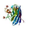
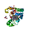
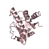
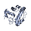
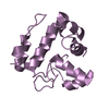
A: Tetrapyrrole-binding protein B: Tetrapyrrole-binding protein C: Tetrapyrrole-binding protein D: Tetrapyrrole-binding protein E: Tetrapyrrole-binding protein F: Tetrapyrrole-binding protein
Mass: 25823.041 Da / Num. of mol.: 6 / Fragment: residues 40-260 Source method: isolated from a genetically manipulated source Source: (gene. exp.) Chlamydomonas reinhardtii (plant) / Gene: GUN4, CHLREDRAFT_205768 / Production host: Escherichia coli (E. coli) / References: UniProt: A8I5N5
-
Experimental details
-
Experiment
Experiment
Method: X-RAY DIFFRACTION
-
Sample preparation
Crystal
Density Matthews: 2.64 Å3/Da / Density % sol: 53 % / Description: very small hexagonal rods
Crystal grow
Temperature: 293 K / Method: vapor diffusion, sitting drop / pH: 8 Details: 1:1 ratio of protein (35 mg/mL in 20 mM Tricine (pH 8.0), 2 mM beta-mercaptoethanol) to well solution (1.0 M ammonium citrate (pH 7.0), 0.1 M Bis Tris Propane (pH 7.0). Crystals only ...Details: 1:1 ratio of protein (35 mg/mL in 20 mM Tricine (pH 8.0), 2 mM beta-mercaptoethanol) to well solution (1.0 M ammonium citrate (pH 7.0), 0.1 M Bis Tris Propane (pH 7.0). Crystals only appeared after 44 weeks, likely the time taken for proteolysis, resulting in the N- and C-terminal truncations PH range: 7.0-8.0
-
Data collection
Diffraction
Mean temperature: 100 K
Diffraction source
Source: SYNCHROTRON / Site: Australian Synchrotron / Beamline: MX2 / Wavelength: 0.9537 Å
Resolution: 3.5→44.5 Å / Cor.coef. Fo:Fc: 0.879 / Cor.coef. Fo:Fc free: 0.813 / SU B: 43.036 / SU ML: 0.663 / Cross valid method: THROUGHOUT / ESU R Free: 0.77 / Stereochemistry target values: MAXIMUM LIKELIHOOD / Details: HYDROGENS HAVE BEEN ADDED IN THE RIDING POSITIONS
Rfactor
Num. reflection
% reflection
Selection details
Rfree
0.31315
753
5.4 %
RANDOM
Rwork
0.25602
-
-
-
obs
0.25907
13187
99.05 %
-
Solvent computation
Ion probe radii: 0.8 Å / Shrinkage radii: 0.8 Å / VDW probe radii: 1.2 Å / Solvent model: MASK
 Movie
Movie Controller


 Open data
 Basic information
 Components
 Keywords
 PLANT PROTEIN
 Function and homology information

 Authors
 Citation
 Structure visualization
 Downloads & links
 Other downloads


 PDBj
PDBj Assembly


 Sample preparation
 / Beamline: MX2 / Wavelength: 0.9537 Å
 Processing