 Movie
Movie Controller


+ Open data

- Basic information
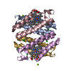
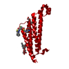
| Entry | Database: PDB / ID: 4yk5 | ||||||
|---|---|---|---|---|---|---|---|
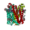
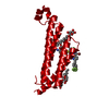
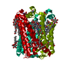
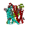
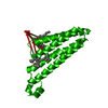
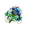
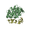
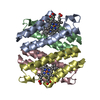
| Title | Crystal Structures of mPGES-1 Inhibitor Complexes | ||||||
 Components Components | Prostaglandin E synthase | ||||||
 Keywords Keywords | ISOMERASE/ISOMERASE INHIBITOR / Inflammation / ISOMERASE-ISOMERASE INHIBITOR complex | ||||||
| Function / homology |  Function and homology information Function and homology informationregulation of fever generation / prostaglandin-E synthase / prostaglandin-E synthase activity / prostaglandin-D synthase activity / positive regulation of prostaglandin secretion / glutathione binding / Synthesis of Prostaglandins (PG) and Thromboxanes (TX) / glutathione peroxidase activity / prostaglandin biosynthetic process / prostaglandin metabolic process ...regulation of fever generation / prostaglandin-E synthase / prostaglandin-E synthase activity / prostaglandin-D synthase activity / positive regulation of prostaglandin secretion / glutathione binding / Synthesis of Prostaglandins (PG) and Thromboxanes (TX) / glutathione peroxidase activity / prostaglandin biosynthetic process / prostaglandin metabolic process / nuclear envelope lumen / glutathione transferase / glutathione transferase activity / Oxidoreductases; Acting on a peroxide as acceptor; Peroxidases / sensory perception of pain / regulation of inflammatory response / cell population proliferation / negative regulation of cell population proliferation / endoplasmic reticulum membrane / perinuclear region of cytoplasm / signal transduction / membraneSimilarity search - Function | ||||||
| Biological species |  Homo sapiens (human) Homo sapiens (human) | ||||||
| Method | X-RAY DIFFRACTION / SYNCHROTRON / Resolution: 1.42 Å | ||||||
 Authors Authors | Luz, J.G. / Antonysamy, S. / Kuklish, S.L. / Fisher, M.J. | ||||||
 Citation Citation | Journal: J.Med.Chem. / Year: 2015 Title: Crystal Structures of mPGES-1 Inhibitor Complexes Form a Basis for the Rational Design of Potent Analgesic and Anti-Inflammatory Therapeutics. Authors: Luz, J.G. / Antonysamy, S. / Kuklish, S.L. / Condon, B. / Lee, M.R. / Allison, D. / Yu, X.P. / Chandrasekhar, S. / Backer, R. / Zhang, A. / Russell, M. / Chang, S.S. / Harvey, A. / Sloan, A.V. / Fisher, M.J. | ||||||
| History |
|
- Structure visualization
| Structure viewer | Molecule: MolmilJmol/JSmol |
|---|
- Downloads & links
-Download
| PDBx/mmCIF format | 4yk5.cif.gz | 85 KB | Display | PDBx/mmCIF format |
|---|---|---|---|---|
| PDB format | pdb4yk5.ent.gz | 61.4 KB | Display | PDB format |
| PDBx/mmJSON format | 4yk5.json.gz | Tree view | PDBx/mmJSON format | |
| Others |  Other downloads Other downloads |
-Validation report
| Arichive directory | https://data.pdbj.org/pub/pdb/validation_reports/yk/4yk5ftp://data.pdbj.org/pub/pdb/validation_reports/yk/4yk5 | HTTPS FTP |
|---|
-Related structure data


-Links
 PDBj
PDBj


- Assembly
| Deposited unit | 
| ||||||||||||
|---|---|---|---|---|---|---|---|---|---|---|---|---|---|
| 1 | 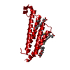
| ||||||||||||
| Unit cell |
| ||||||||||||
| Components on special symmetry positions |
|
-Components
-Protein , 1 types, 1 molecules A
| #1: Protein | / Microsomal glutathione S-transferase 1-like 1 / MGST1-L1 / Microsomal prostaglandin E synthase 1 / ...Microsomal glutathione S-transferase 1-like 1 / MGST1-L1 / Microsomal prostaglandin E synthase 1 / MPGES-1 / p53-induced gene 12 protein Mass: 17249.414 Da / Num. of mol.: 1 Source method: isolated from a genetically manipulated source Source: (gene. exp.) Homo sapiens (human) / Gene: PTGES, MGST1L1, MPGES1, PGES, PIG12 / Production host:   Spodoptera frugiperda (fall armyworm) / References: UniProt: O14684, prostaglandin-E synthase Spodoptera frugiperda (fall armyworm) / References: UniProt: O14684, prostaglandin-E synthase |
|---|
-Sugars , 2 types, 2 molecules 

| #5: Sugar | ChemComp-JZR /  Type: D-saccharide / Mass: 264.315 Da / Num. of mol.: 1 Type: D-saccharide / Mass: 264.315 Da / Num. of mol.: 1Source method: isolated from a genetically manipulated source Formula: C12H24O6 |
|---|---|
| #6: Sugar | ChemComp-BOG / Octyl glucoside Type: D-saccharide / Mass: 292.369 Da / Num. of mol.: 1 Type: D-saccharide / Mass: 292.369 Da / Num. of mol.: 1Source method: isolated from a genetically manipulated source Formula: C14H28O6 / Comment: detergent*YM |
-Non-polymers , 5 types, 150 molecules 


| #2: Chemical | ChemComp-4DV /  Mass: 540.067 Da / Num. of mol.: 1 / Source method: obtained synthetically / Formula: C34H31ClFNO2 Mass: 540.067 Da / Num. of mol.: 1 / Source method: obtained synthetically / Formula: C34H31ClFNO2 |
|---|---|
| #3: Chemical | ChemComp-PG0 / 2-(2-Methoxyethoxy)ethanol Mass: 120.147 Da / Num. of mol.: 1 / Source method: obtained synthetically / Formula: C5H12O3 / Comment: inhibitor, precipitant*YM Mass: 120.147 Da / Num. of mol.: 1 / Source method: obtained synthetically / Formula: C5H12O3 / Comment: inhibitor, precipitant*YM |
| #4: Chemical | ChemComp-GSH / Glutathione Mass: 307.323 Da / Num. of mol.: 1 Mass: 307.323 Da / Num. of mol.: 1Source method: isolated from a genetically manipulated source Formula: C10H17N3O6S |
| #7: Chemical | ChemComp-PG4 / Polyethylene glycol Mass: 194.226 Da / Num. of mol.: 1 / Source method: obtained synthetically / Formula: C8H18O5 / Comment: precipitant*YM Mass: 194.226 Da / Num. of mol.: 1 / Source method: obtained synthetically / Formula: C8H18O5 / Comment: precipitant*YM |
| #8: Water | ChemComp-HOH / WaterMass: 18.015 Da / Num. of mol.: 146 / Source method: isolated from a natural source / Formula: H2O |
-Experimental details
-Experiment
| Experiment | Method: X-RAY DIFFRACTION |
|---|
- Sample preparation
| Crystal | Density Matthews: 4.05 Å3/Da / Density % sol: 69.63 % |
|---|---|
| Crystal grow | Temperature: 294.15 K / Method: vapor diffusion, sitting drop / Details: 100mM Tris HCl pH 8.5 + 32.5% PEG 1K |
-Data collection
| Diffraction | Mean temperature: 195 K |
|---|---|
| Diffraction source | Source: SYNCHROTRON / Site: APS  / Beamline: 31-ID / Wavelength: 0.97929 Å / Beamline: 31-ID / Wavelength: 0.97929 Å |
| Detector | Type: RAYONIX MX225HE / Detector: CCD / Date: Apr 2, 2009 |
| Radiation | Protocol: SINGLE WAVELENGTH / Monochromatic (M) / Laue (L): M / Scattering type: x-ray |
| Radiation wavelength | Wavelength: 0.97929 Å / Relative weight: 1 |
| Reflection | Resolution: 1.42→32 Å / Num. obs: 50979 / % possible obs: 99.3 % / Redundancy: 5.4 % / Net I/σ(I): 14.9 |
- Processing
| Software |
| ||||||||||||||||||||||||||||||||||||||||||||||||||||||||||||||||||||||||||||||||||||||||||||||||||||||||||||||||||||||||||||||||||||||||||||||||||||||||||||||||||||||||||||||||||||||
|---|---|---|---|---|---|---|---|---|---|---|---|---|---|---|---|---|---|---|---|---|---|---|---|---|---|---|---|---|---|---|---|---|---|---|---|---|---|---|---|---|---|---|---|---|---|---|---|---|---|---|---|---|---|---|---|---|---|---|---|---|---|---|---|---|---|---|---|---|---|---|---|---|---|---|---|---|---|---|---|---|---|---|---|---|---|---|---|---|---|---|---|---|---|---|---|---|---|---|---|---|---|---|---|---|---|---|---|---|---|---|---|---|---|---|---|---|---|---|---|---|---|---|---|---|---|---|---|---|---|---|---|---|---|---|---|---|---|---|---|---|---|---|---|---|---|---|---|---|---|---|---|---|---|---|---|---|---|---|---|---|---|---|---|---|---|---|---|---|---|---|---|---|---|---|---|---|---|---|---|---|---|---|---|
| Refinement | Resolution: 1.42→30 Å / Cor.coef. Fo:Fc: 0.964 / Cor.coef. Fo:Fc free: 0.964 / SU B: 1.274 / SU ML: 0.023 / Cross valid method: THROUGHOUT / ESU R: 0.047 / ESU R Free: 0.042 / Stereochemistry target values: MAXIMUM LIKELIHOOD
| ||||||||||||||||||||||||||||||||||||||||||||||||||||||||||||||||||||||||||||||||||||||||||||||||||||||||||||||||||||||||||||||||||||||||||||||||||||||||||||||||||||||||||||||||||||||
| Solvent computation | Ion probe radii: 0.8 Å / Shrinkage radii: 0.8 Å / VDW probe radii: 1.2 Å / Solvent model: MASK | ||||||||||||||||||||||||||||||||||||||||||||||||||||||||||||||||||||||||||||||||||||||||||||||||||||||||||||||||||||||||||||||||||||||||||||||||||||||||||||||||||||||||||||||||||||||
| Displacement parameters | Biso mean: 19.89 Å2
| ||||||||||||||||||||||||||||||||||||||||||||||||||||||||||||||||||||||||||||||||||||||||||||||||||||||||||||||||||||||||||||||||||||||||||||||||||||||||||||||||||||||||||||||||||||||
| Refinement step | Cycle: LAST / Resolution: 1.42→30 Å
| ||||||||||||||||||||||||||||||||||||||||||||||||||||||||||||||||||||||||||||||||||||||||||||||||||||||||||||||||||||||||||||||||||||||||||||||||||||||||||||||||||||||||||||||||||||||
| Refine LS restraints |
|