 Movie
Movie Controller

[English] 日本語
 Yorodumi
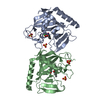
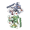
- PDB-4yjz: Human antibody H2526 in complex with influenza hemagglutinin H1 S... -
+ Open data

- Basic information
| Entry | Database: PDB / ID: 4yjz | ||||||
|---|---|---|---|---|---|---|---|
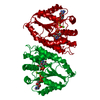
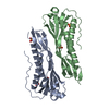
| Title | Human antibody H2526 in complex with influenza hemagglutinin H1 Solomon Islands/03/2006 | ||||||
 Components Components |
| ||||||
 Keywords Keywords |  Viral protein/Immune system / influenza / antibody / complex / hemagglutinin / Viral protein-Immune system complex Viral protein/Immune system / influenza / antibody / complex / hemagglutinin / Viral protein-Immune system complex | ||||||
| Function / homology |  Function and homology informationviral budding from plasma membrane / clathrin-dependent endocytosis of virus by host cell / host cell surface receptor binding / apical plasma membrane / fusion of virus membrane with host plasma membrane / fusion of virus membrane with host endosome membrane / viral envelope / virion attachment to host cell / host cell plasma membrane / virion membrane Function and homology informationviral budding from plasma membrane / clathrin-dependent endocytosis of virus by host cell / host cell surface receptor binding / apical plasma membrane / fusion of virus membrane with host plasma membrane / fusion of virus membrane with host endosome membrane / viral envelope / virion attachment to host cell / host cell plasma membrane / virion membraneSimilarity search - Function | ||||||
| Biological species |   Influenza A virus Influenza A virus Homo sapiens (human) Homo sapiens (human) | ||||||
| Method | X-RAY DIFFRACTION / SYNCHROTRON / MOLECULAR REPLACEMENT / molecular replacement / Resolution: 2.72 Å | ||||||
 Authors Authors | Schmidt, A.G. / Harrison, S.C. | ||||||
 Citation Citation | Journal: Cell / Year: 2015 Title: Viral receptor-binding site antibodies with diverse germline origins. Authors: Schmidt, A.G. / Therkelsen, M.D. / Stewart, S. / Kepler, T.B. / Liao, H.X. / Moody, M.A. / Haynes, B.F. / Harrison, S.C. | ||||||
| History |
|
- Structure visualization
| Structure viewer | Molecule: MolmilJmol/JSmol |
|---|
- Downloads & links
-Download
| PDBx/mmCIF format | 4yjz.cif.gz | 105 KB | Display | PDBx/mmCIF format |
|---|---|---|---|---|
| PDB format | pdb4yjz.ent.gz | 78.2 KB | Display | PDB format |
| PDBx/mmJSON format | 4yjz.json.gz | Tree view | PDBx/mmJSON format | |
| Others |  Other downloads Other downloads |
-Validation report
| Arichive directory | https://data.pdbj.org/pub/pdb/validation_reports/yj/4yjzftp://data.pdbj.org/pub/pdb/validation_reports/yj/4yjz | HTTPS FTP |
|---|
-Related structure data
| Related structure data |  4yk4C  3gbnS  4hkxS C: citing same article ( S: Starting model for refinement |
|---|---|
| Similar structure data |


-Links
 PDBj
PDBj


- Assembly
| Deposited unit | 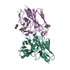
| ||||||||
|---|---|---|---|---|---|---|---|---|---|
| 1 |
| ||||||||
| Unit cell |
|
-Components
| #1: Protein | Mass: 25109.988 Da / Num. of mol.: 1 Source method: isolated from a genetically manipulated source Source: (gene. exp.) Influenza A virus / Strain: A/Solomon Islands/3/2006(H1N1) / Gene: HA / Production host:  Trichoplusia ni (cabbage looper) / References: UniProt: A7UPX0 Trichoplusia ni (cabbage looper) / References: UniProt: A7UPX0 | ||||
|---|---|---|---|---|---|
| #2: Antibody | Mass: 27967.621 Da / Num. of mol.: 1 Source method: isolated from a genetically manipulated source Source: (gene. exp.) Homo sapiens (human) / Production host: Mammalia (mammals) | ||||
| #3: Sugar | N-Acetylglucosamine  Type: D-saccharide, beta linking / Mass: 221.208 Da / Num. of mol.: 3 Type: D-saccharide, beta linking / Mass: 221.208 Da / Num. of mol.: 3Source method: isolated from a genetically manipulated source Formula: C8H15NO6 #4: Water | ChemComp-HOH / | Water Mass: 18.015 Da / Num. of mol.: 48 / Source method: isolated from a natural source / Formula: H2O Mass: 18.015 Da / Num. of mol.: 48 / Source method: isolated from a natural source / Formula: H2OSequence details | Chain L in this structure is a single-chain variable fragment (scFv) which includes the variable ...Chain L in this structure is a single-chain variable fragment (scFv) which includes the variable heavy and variable light domains of an antibody with a linker in-between (GGGGGGSGGG | |
-Experimental details
-Experiment
| Experiment | Method: X-RAY DIFFRACTION / Number of used crystals: 1 |
|---|
- Sample preparation
| Crystal | Density Matthews: 4.05 Å3/Da / Density % sol: 69.6 % |
|---|---|
| Crystal grow | Temperature: 293 K / Method: vapor diffusion, hanging drop / pH: 7 / Details: 0.1M HEPES, 30% PEG 400 and 0.1M ammonium sulfate |
-Data collection
| Diffraction | Mean temperature: 100 K |
|---|---|
| Diffraction source | Source: SYNCHROTRON / Site: ALS  / Beamline: 8.2.2 / Wavelength: 0.99998 Å / Beamline: 8.2.2 / Wavelength: 0.99998 Å |
| Detector | Type: ADSC QUANTUM 315 / Detector: CCD / Date: Feb 6, 2014 |
| Diffraction measurement | Details: 1.00 degrees, 5.0 sec, detector distance 350.01 mm / Method: \w scans |
| Radiation | Protocol: SINGLE WAVELENGTH / Monochromatic (M) / Laue (L): M / Scattering type: x-ray |
| Radiation wavelength | Wavelength: 0.99998 Å / Relative weight: 1 |
| Reflection | Av R equivalents: 0.07 / Number: 135025 |
| Reflection | Resolution: 2.72→50 Å / Num. obs: 23649 / % possible obs: 99.8 % / Observed criterion σ(F): 0 / Observed criterion σ(I): -3 / Redundancy: 5.7 % / Rmerge(I) obs: 0.07 / Rsym value: 0.07 / Net I/av σ(I): 39.699 |
| Reflection shell | Resolution: 2.72→2.77 Å / Redundancy: 5.9 % / Rmerge(I) obs: 0.648 / Mean I/σ(I) obs: 2.892 / Rsym value: 0.648 / % possible all: 100 |
| Cell measurement | Reflection used: 135025 |
-Phasing
| Phasing | Method: molecular replacement |
|---|
- Processing
| Software |
| |||||||||||||||||||||||||||||||||||||||||||||||||||||||||||||||
|---|---|---|---|---|---|---|---|---|---|---|---|---|---|---|---|---|---|---|---|---|---|---|---|---|---|---|---|---|---|---|---|---|---|---|---|---|---|---|---|---|---|---|---|---|---|---|---|---|---|---|---|---|---|---|---|---|---|---|---|---|---|---|---|---|
| Refinement | Method to determine structure: MOLECULAR REPLACEMENT Starting model: 3GBN, 4HKX Resolution: 2.72→39.703 Å / FOM work R set: 0.7241 / SU ML: 0.39 / Cross valid method: FREE R-VALUE / σ(F): 1.34 / Phase error: 32.68 / Stereochemistry target values: ML
| |||||||||||||||||||||||||||||||||||||||||||||||||||||||||||||||
| Solvent computation | Shrinkage radii: 0.9 Å / VDW probe radii: 1.11 Å / Solvent model: FLAT BULK SOLVENT MODEL | |||||||||||||||||||||||||||||||||||||||||||||||||||||||||||||||
| Displacement parameters | Biso max: 182.19 Å2 / Biso mean: 90.78 Å2 / Biso min: 46.68 Å2 | |||||||||||||||||||||||||||||||||||||||||||||||||||||||||||||||
| Refinement step | Cycle: final / Resolution: 2.72→39.703 Å
| |||||||||||||||||||||||||||||||||||||||||||||||||||||||||||||||
| Refine LS restraints |
| |||||||||||||||||||||||||||||||||||||||||||||||||||||||||||||||
| LS refinement shell | Refine-ID: X-RAY DIFFRACTION / Total num. of bins used: 8
|
