 Movie
Movie Controller


+ Open data

- Basic information
| Entry | Database: PDB / ID: 4yj1 | ||||||
|---|---|---|---|---|---|---|---|
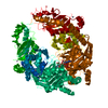
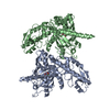
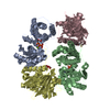
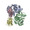
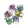
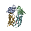
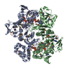
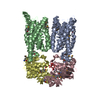
| Title | Crystal structure of T. brucei MRB1590-ADP bound to poly-U RNA | ||||||
 Components Components | Uncharacterized protein | ||||||
 Keywords Keywords |  RNA BINDING PROTEIN / kRNA editing / KASP / MRB1 RNA BINDING PROTEIN / kRNA editing / KASP / MRB1 | ||||||
| Function / homology |  Function and homology information Function and homology informationmitochondrial mRNA processing / mitochondrial RNA processing / cytidine to uridine editing / mRNA stabilization / nucleotide binding / mRNA binding / RNA binding / cytoplasmSimilarity search - Function | ||||||
| Biological species |  Trypanosoma brucei brucei (eukaryote) Trypanosoma brucei brucei (eukaryote) | ||||||
| Method | X-RAY DIFFRACTION / SYNCHROTRON / MOLECULAR REPLACEMENT / Resolution: 2.05 Å | ||||||
 Authors Authors | Schumacher, M.A. | ||||||
 Citation Citation | Journal: Nucleic Acids Res. / Year: 2015 Title: Structures of the T. brucei kRNA editing factor MRB1590 reveal unique RNA-binding pore motif contained within an ABC-ATPase fold. Authors: Shaw, P.L. / McAdams, N.M. / Hast, M.A. / Ammerman, M.L. / Read, L.K. / Schumacher, M.A. | ||||||
| History |
|
- Structure visualization
| Structure viewer | Molecule: MolmilJmol/JSmol |
|---|
- Downloads & links
-Download
| PDBx/mmCIF format | 4yj1.cif.gz | 242.2 KB | Display | PDBx/mmCIF format |
|---|---|---|---|---|
| PDB format | pdb4yj1.ent.gz | 193.6 KB | Display | PDB format |
| PDBx/mmJSON format | 4yj1.json.gz | Tree view | PDBx/mmJSON format | |
| Others |  Other downloads Other downloads |
-Validation report
| Arichive directory | https://data.pdbj.org/pub/pdb/validation_reports/yj/4yj1ftp://data.pdbj.org/pub/pdb/validation_reports/yj/4yj1 | HTTPS FTP |
|---|
-Related structure data


-Links
 PDBj
PDBj- Assembly
| Deposited unit | 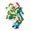
| |||||||||||||||||||||
|---|---|---|---|---|---|---|---|---|---|---|---|---|---|---|---|---|---|---|---|---|---|---|
| 1 | 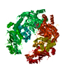
| |||||||||||||||||||||
| Unit cell |
| |||||||||||||||||||||
| Components on special symmetry positions |
|
-Components
| #1: Protein | Mass: 67054.977 Da / Num. of mol.: 1 / Fragment: UNP residues 50-665 Source method: isolated from a genetically manipulated source Source: (gene. exp.) Trypanosoma brucei brucei (strain 927/4 GUTat10.1) (eukaryote)Strain: 927/4 GUTat10.1 / Gene: Tb927.3.1590 / Production host:  Escherichia coli (E. coli) / References: UniProt: Q57ZF2 Escherichia coli (E. coli) / References: UniProt: Q57ZF2 | ||
|---|---|---|---|
| #2: Chemical | ChemComp-ADP / Adenosine diphosphate  Mass: 427.201 Da / Num. of mol.: 1 / Source method: obtained synthetically / Formula: C10H15N5O10P2 / Comment: ADP, energy-carrying molecule*YM Mass: 427.201 Da / Num. of mol.: 1 / Source method: obtained synthetically / Formula: C10H15N5O10P2 / Comment: ADP, energy-carrying molecule*YM | ||
| #3: Chemical |   Mass: 24.305 Da / Num. of mol.: 2 / Source method: obtained synthetically / Formula: Mg Mass: 24.305 Da / Num. of mol.: 2 / Source method: obtained synthetically / Formula: Mg#4: Water | ChemComp-HOH / | Water Mass: 18.015 Da / Num. of mol.: 521 / Source method: isolated from a natural source / Formula: H2O Mass: 18.015 Da / Num. of mol.: 521 / Source method: isolated from a natural source / Formula: H2O |
-Experimental details
-Experiment
| Experiment | Method: X-RAY DIFFRACTION / Number of used crystals: 1 |
|---|
- Sample preparation
| Crystal | Density Matthews: 4.33 Å3/Da / Density % sol: 71.61 % |
|---|---|
| Crystal grow | Temperature: 298 K / Method: vapor diffusion, hanging drop / Details: PEG 3350, magnesium chloride |
-Data collection
| Diffraction | Mean temperature: 100 K |
|---|---|
| Diffraction source | Source: SYNCHROTRON / Site: ALS  / Beamline: 8.3.1 / Wavelength: 1 Å / Beamline: 8.3.1 / Wavelength: 1 Å |
| Detector | Type: ADSC QUANTUM 315r / Detector: CCD / Date: Mar 4, 2013 |
| Radiation | Protocol: SINGLE WAVELENGTH / Monochromatic (M) / Laue (L): M / Scattering type: x-ray |
| Radiation wavelength | Wavelength: 1 Å / Relative weight: 1 |
| Reflection | Resolution: 2.05→41.8 Å / Num. obs: 70357 / % possible obs: 95.6 % / Redundancy: 4 % / Net I/σ(I): 12 |
- Processing
| Software |
| |||||||||||||||||||||||||||||||||||||||||||||||||||||||||||||||||||||||||||||
|---|---|---|---|---|---|---|---|---|---|---|---|---|---|---|---|---|---|---|---|---|---|---|---|---|---|---|---|---|---|---|---|---|---|---|---|---|---|---|---|---|---|---|---|---|---|---|---|---|---|---|---|---|---|---|---|---|---|---|---|---|---|---|---|---|---|---|---|---|---|---|---|---|---|---|---|---|---|---|
| Refinement | Method to determine structure: MOLECULAR REPLACEMENT / Resolution: 2.05→41.8 Å / FOM work R set: 0.8779 / SU ML: 0.23 / Cross valid method: THROUGHOUT / σ(F): 0 / Phase error: 19 / Stereochemistry target values: ML
| |||||||||||||||||||||||||||||||||||||||||||||||||||||||||||||||||||||||||||||
| Solvent computation | Shrinkage radii: 0.77 Å / VDW probe radii: 0.9 Å / Solvent model: FLAT BULK SOLVENT MODEL / Bsol: 42.931 Å2 / ksol: 0.375 e/Å3 | |||||||||||||||||||||||||||||||||||||||||||||||||||||||||||||||||||||||||||||
| Displacement parameters | Biso max: 148 Å2 / Biso mean: 32.1 Å2 / Biso min: 11 Å2
| |||||||||||||||||||||||||||||||||||||||||||||||||||||||||||||||||||||||||||||
| Refinement step | Cycle: final / Resolution: 2.05→41.8 Å
| |||||||||||||||||||||||||||||||||||||||||||||||||||||||||||||||||||||||||||||
| Refine LS restraints |
| |||||||||||||||||||||||||||||||||||||||||||||||||||||||||||||||||||||||||||||
| LS refinement shell | Refine-ID: X-RAY DIFFRACTION / Total num. of bins used: 10
|
