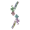
 Movie
Movie Controller


+ Open data

- Basic information
| Entry | Database: PDB / ID: 4yfg | ||||||
|---|---|---|---|---|---|---|---|
| Title | Crystal structure of PTP delta meA3/meB minus variant Ig1-Fn1 | ||||||
 Components Components | Receptor-type tyrosine-protein phosphatase delta | ||||||
 Keywords Keywords |  HYDROLASE / synapse organizer HYDROLASE / synapse organizer | ||||||
| Function / homology |  Function and homology information Function and homology informationReceptor-type tyrosine-protein phosphatases / trans-synaptic signaling / Synaptic adhesion-like molecules / trans-synaptic signaling by trans-synaptic complex / presynaptic membrane assembly / synaptic membrane adhesion / regulation of postsynaptic density assembly / negative regulation of receptor signaling pathway via JAK-STAT / positive regulation of synapse assembly / positive regulation of dendrite morphogenesis ...Receptor-type tyrosine-protein phosphatases / trans-synaptic signaling / Synaptic adhesion-like molecules / trans-synaptic signaling by trans-synaptic complex / presynaptic membrane assembly / synaptic membrane adhesion / regulation of postsynaptic density assembly / negative regulation of receptor signaling pathway via JAK-STAT / positive regulation of synapse assembly / positive regulation of dendrite morphogenesis / heterophilic cell-cell adhesion via plasma membrane cell adhesion molecules / regulation of immune response / dephosphorylation / cell adhesion molecule binding / hippocampal mossy fiber to CA3 synapse / protein-tyrosine-phosphatase / protein tyrosine phosphatase activity / Schaffer collateral - CA1 synapse / modulation of chemical synaptic transmission / neuron differentiation / presynaptic membrane / receptor complex / signaling receptor binding / glutamatergic synapseSimilarity search - Function | ||||||
| Biological species |  Mus musculus (house mouse) Mus musculus (house mouse) | ||||||
| Method | X-RAY DIFFRACTION / SYNCHROTRON / MOLECULAR REPLACEMENT / Resolution: 3.491 Å | ||||||
 Authors Authors | Yamagata, A. / Fukai, S. | ||||||
 Citation Citation | Journal: Nat Commun / Year: 2015 Title: Mechanisms of splicing-dependent trans-synaptic adhesion by PTP delta-IL1RAPL1/IL-1RAcP for synaptic differentiation. Authors: Yamagata, A. / Yoshida, T. / Sato, Y. / Goto-Ito, S. / Uemura, T. / Maeda, A. / Shiroshima, T. / Iwasawa-Okamoto, S. / Mori, H. / Mishina, M. / Fukai, S. | ||||||
| History |
|
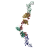
- Structure visualization
| Structure viewer | Molecule: MolmilJmol/JSmol |
|---|
- Downloads & links
-Download
| PDBx/mmCIF format | 4yfg.cif.gz | 384.7 KB | Display | PDBx/mmCIF format |
|---|---|---|---|---|
| PDB format | pdb4yfg.ent.gz | 315.4 KB | Display | PDB format |
| PDBx/mmJSON format | 4yfg.json.gz | Tree view | PDBx/mmJSON format | |
| Others |  Other downloads Other downloads |
-Validation report
| Arichive directory | https://data.pdbj.org/pub/pdb/validation_reports/yf/4yfgftp://data.pdbj.org/pub/pdb/validation_reports/yf/4yfg | HTTPS FTP |
|---|
-Related structure data
| Related structure data | 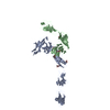 4yfdC 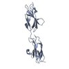 4yfeSC 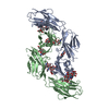 4yh6C 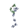 4yh7C 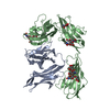 5y32C 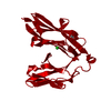 2yd6S  2yd9S C: citing same article ( S: Starting model for refinement |
|---|---|
| Similar structure data |

-Links
 PDBj
PDBj


- Assembly
| Deposited unit | 
| ||||||||
|---|---|---|---|---|---|---|---|---|---|
| 1 |
| ||||||||
| Unit cell |
|
-Components
| #1: Protein | Mass: 53759.250 Da / Num. of mol.: 2 / Fragment: UNP RESIDUES 21-501 Source method: isolated from a genetically manipulated source Source: (gene. exp.) Mus musculus (house mouse) / Gene: Ptprd / Cell line (production host): Freestyle 293-F / Production host:  Homo sapiens (human) / References: UniProt: Q64487, protein-tyrosine-phosphatase Homo sapiens (human) / References: UniProt: Q64487, protein-tyrosine-phosphatase#2: Sugar | ChemComp-NAG / N-Acetylglucosamine  Type: D-saccharide, beta linking / Mass: 221.208 Da / Num. of mol.: 4 Type: D-saccharide, beta linking / Mass: 221.208 Da / Num. of mol.: 4Source method: isolated from a genetically manipulated source Formula: C8H15NO6 Sequence details | SEQUENCE OF THE PROTEIN WAS BASED ON ISOFORM 9 OF DATABASE UNP Q64487 | |
|---|
-Experimental details
-Experiment
| Experiment | Method: X-RAY DIFFRACTION |
|---|
- Sample preparation
| Crystal | Density Matthews: 3.96 Å3/Da / Density % sol: 68.92 % |
|---|---|
| Crystal grow | Temperature: 293 K / Method: vapor diffusion, sitting drop Details: 10% polyethylene glycol (PEG) 3350, 0.1M Ammonium iodide |
-Data collection
| Diffraction | Mean temperature: 100 K |
|---|---|
| Diffraction source | Source: SYNCHROTRON / Site: SPring-8  / Beamline: BL41XU / Wavelength: 1 Å / Beamline: BL41XU / Wavelength: 1 Å |
| Detector | Type: RAYONIX MX225HE / Detector: CCD / Date: Jan 31, 2014 |
| Radiation | Protocol: SINGLE WAVELENGTH / Monochromatic (M) / Laue (L): M / Scattering type: x-ray |
| Radiation wavelength | Wavelength: 1 Å / Relative weight: 1 |
| Reflection | Resolution: 3.491→50 Å / Num. obs: 18642 / % possible obs: 91.4 % / Redundancy: 2.5 % / Rsym value: 0.189 / Net I/σ(I): 4.8 |
| Reflection shell | Resolution: 3.5→3.56 Å / Redundancy: 1.9 % / Rmerge(I) obs: 0.491 / Mean I/σ(I) obs: 1.9 / % possible all: 84.2 |
- Processing
| Software |
| ||||||||||||||||||||||||||||||||||||||||||||||||||||||||
|---|---|---|---|---|---|---|---|---|---|---|---|---|---|---|---|---|---|---|---|---|---|---|---|---|---|---|---|---|---|---|---|---|---|---|---|---|---|---|---|---|---|---|---|---|---|---|---|---|---|---|---|---|---|---|---|---|---|
| Refinement | Method to determine structure: MOLECULAR REPLACEMENT Starting model: 2YD6, 2YD9, 4YFE Resolution: 3.491→48.406 Å / Cross valid method: FREE R-VALUE / σ(F): 1.97 / Phase error: 37.03 / Stereochemistry target values: ML
| ||||||||||||||||||||||||||||||||||||||||||||||||||||||||
| Solvent computation | Shrinkage radii: 0.9 Å / VDW probe radii: 1.11 Å / Solvent model: FLAT BULK SOLVENT MODEL | ||||||||||||||||||||||||||||||||||||||||||||||||||||||||
| Refinement step | Cycle: LAST / Resolution: 3.491→48.406 Å
| ||||||||||||||||||||||||||||||||||||||||||||||||||||||||
| Refine LS restraints |
| ||||||||||||||||||||||||||||||||||||||||||||||||||||||||
| LS refinement shell | Refine-ID: X-RAY DIFFRACTION
| ||||||||||||||||||||||||||||||||||||||||||||||||||||||||
| Refinement TLS params. | Method: refined / Origin x: 17.2084 Å / Origin y: 9.9145 Å / Origin z: -7.2971 Å
| ||||||||||||||||||||||||||||||||||||||||||||||||||||||||
| Refinement TLS group | Selection details: all |
