 Movie
Movie Controller


+ Open data

- Basic information
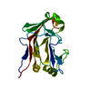
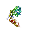
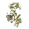
| Entry | Database: PDB / ID: 4yep | |||||||||
|---|---|---|---|---|---|---|---|---|---|---|
| Title | L4b Domain of Human Laminin alpha-2 | |||||||||
 Components Components | Laminin subunit alpha-2 | |||||||||
 Keywords Keywords | SUGAR BINDING PROTEIN / carbohydrate binding fold / laminin / extracellular matrix / ephrin receptor | |||||||||
| Function / homology |  Function and homology information Function and homology informationregulation of basement membrane organization / Schwann cell differentiation / positive regulation of synaptic transmission, cholinergic / positive regulation of integrin-mediated signaling pathway / Laminin interactions / EGR2 and SOX10-mediated initiation of Schwann cell myelination / protein complex involved in cell-matrix adhesion / muscle organ development / MET activates PTK2 signaling / maintenance of blood-brain barrier ...regulation of basement membrane organization / Schwann cell differentiation / positive regulation of synaptic transmission, cholinergic / positive regulation of integrin-mediated signaling pathway / Laminin interactions / EGR2 and SOX10-mediated initiation of Schwann cell myelination / protein complex involved in cell-matrix adhesion / muscle organ development / MET activates PTK2 signaling / maintenance of blood-brain barrier / positive regulation of muscle cell differentiation / regulation of embryonic development / positive regulation of cell adhesion / Non-integrin membrane-ECM interactions / basement membrane / ECM proteoglycans / synaptic cleft / regulation of cell migration / axon guidance / neuromuscular junction / sarcolemma / collagen-containing extracellular matrix / dendritic spine / cell adhesion / signaling receptor binding / structural molecule activity / extracellular regionSimilarity search - Function | |||||||||
| Biological species |  Homo sapiens (human) Homo sapiens (human) | |||||||||
| Method | X-RAY DIFFRACTION / SYNCHROTRON / Resolution: 1.19 Å | |||||||||
 Authors Authors | Toot, M. / Gat, Y. / Fass, D. | |||||||||
| Funding support |  Israel, 2items Israel, 2items
| |||||||||
 Citation Citation | Journal: Febs J. / Year: 2015 Title: Laminin L4 domain structure resembles adhesion modules in ephrin receptor and other transmembrane glycoproteins. Authors: Moran, T. / Gat, Y. / Fass, D. | |||||||||
| History |
|
- Structure visualization
| Structure viewer | Molecule: MolmilJmol/JSmol |
|---|
- Downloads & links
-Download
| PDBx/mmCIF format | 4yep.cif.gz | 267.7 KB | Display | PDBx/mmCIF format |
|---|---|---|---|---|
| PDB format | pdb4yep.ent.gz | 221.6 KB | Display | PDB format |
| PDBx/mmJSON format | 4yep.json.gz | Tree view | PDBx/mmJSON format | |
| Others |  Other downloads Other downloads |
-Validation report
| Arichive directory | https://data.pdbj.org/pub/pdb/validation_reports/ye/4yepftp://data.pdbj.org/pub/pdb/validation_reports/ye/4yep | HTTPS FTP |
|---|
-Related structure data


-Links
 PDBj
PDBj


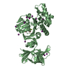
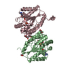
- Assembly
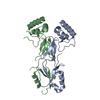
| Deposited unit | 
| |||||||||
|---|---|---|---|---|---|---|---|---|---|---|
| 1 |
| |||||||||
| Unit cell |
| |||||||||
| Components on special symmetry positions |
|
-Components
| #1: Protein | / Laminin M chain / Laminin-12 subunit alpha / Laminin-2 subunit alpha / Laminin-4 subunit alpha / ...Laminin M chain / Laminin-12 subunit alpha / Laminin-2 subunit alpha / Laminin-4 subunit alpha / Merosin heavy chain Mass: 21715.029 Da / Num. of mol.: 2 / Fragment: L4b domain, UNP residues 1181-1362 Source method: isolated from a genetically manipulated source Source: (gene. exp.) Homo sapiens (human) / Gene: LAMA2, LAMM / Plasmid: pMal-c2 / Production host:  Escherichia coli BL21(DE3) (bacteria) / References: UniProt: P24043 Escherichia coli BL21(DE3) (bacteria) / References: UniProt: P24043#2: Chemical | ChemComp-EDO / Ethylene glycol  Mass: 62.068 Da / Num. of mol.: 15 / Source method: obtained synthetically / Formula: C2H6O2 Mass: 62.068 Da / Num. of mol.: 15 / Source method: obtained synthetically / Formula: C2H6O2#3: Water | ChemComp-HOH / | Water Mass: 18.015 Da / Num. of mol.: 480 / Source method: isolated from a natural source / Formula: H2O Mass: 18.015 Da / Num. of mol.: 480 / Source method: isolated from a natural source / Formula: H2O |
|---|
-Experimental details
-Experiment
| Experiment | Method: X-RAY DIFFRACTION |
|---|
- Sample preparation
| Crystal | Density Matthews: 2.82 Å3/Da / Density % sol: 56 % |
|---|---|
| Crystal grow | Temperature: 293 K / Method: vapor diffusion, hanging drop / pH: 8 Details: 15% PEG 3350, 20% ethylene glycol, 50 mM sodium phosphate pH 8.0 PH range: 8 |
-Data collection
| Diffraction | Mean temperature: 100 K |
|---|---|
| Diffraction source | Source: SYNCHROTRON / Site: ESRF  / Beamline: ID29 / Wavelength: 0.919 Å / Beamline: ID29 / Wavelength: 0.919 Å |
| Detector | Type: DECTRIS PILATUS 6M / Detector: PIXEL / Date: Jun 16, 2014 |
| Radiation | Protocol: SINGLE WAVELENGTH / Monochromatic (M) / Laue (L): M / Scattering type: x-ray |
| Radiation wavelength | Wavelength: 0.919 Å / Relative weight: 1 |
| Reflection | Resolution: 1.19→52.85 Å / Num. obs: 110147 / % possible obs: 99.9 % / Redundancy: 13 % / Net I/σ(I): 12.2 |
| Reflection shell | Resolution: 1.19→1.232 Å / Redundancy: 6.6 % / Mean I/σ(I) obs: 2 / % possible all: 99.7 |
- Processing
| Software |
| |||||||||||||||||||||||||||||||||||||||||||||||||||||||||||||||||||||||||||||||||||||||||||||||||||||||||||||||||||||||||||||||||||||||||||||||||||||||||||||||||||||||||||||||||||||||||||||||||||||||||||||||||||||||||
|---|---|---|---|---|---|---|---|---|---|---|---|---|---|---|---|---|---|---|---|---|---|---|---|---|---|---|---|---|---|---|---|---|---|---|---|---|---|---|---|---|---|---|---|---|---|---|---|---|---|---|---|---|---|---|---|---|---|---|---|---|---|---|---|---|---|---|---|---|---|---|---|---|---|---|---|---|---|---|---|---|---|---|---|---|---|---|---|---|---|---|---|---|---|---|---|---|---|---|---|---|---|---|---|---|---|---|---|---|---|---|---|---|---|---|---|---|---|---|---|---|---|---|---|---|---|---|---|---|---|---|---|---|---|---|---|---|---|---|---|---|---|---|---|---|---|---|---|---|---|---|---|---|---|---|---|---|---|---|---|---|---|---|---|---|---|---|---|---|---|---|---|---|---|---|---|---|---|---|---|---|---|---|---|---|---|---|---|---|---|---|---|---|---|---|---|---|---|---|---|---|---|---|---|---|---|---|---|---|---|---|---|---|---|---|---|---|---|---|
| Refinement | Resolution: 1.19→52.846 Å / SU ML: 0.11 / Cross valid method: FREE R-VALUE / σ(F): 1.34 / Phase error: 15.27 / Stereochemistry target values: ML
| |||||||||||||||||||||||||||||||||||||||||||||||||||||||||||||||||||||||||||||||||||||||||||||||||||||||||||||||||||||||||||||||||||||||||||||||||||||||||||||||||||||||||||||||||||||||||||||||||||||||||||||||||||||||||
| Solvent computation | Shrinkage radii: 0.9 Å / VDW probe radii: 1.11 Å / Solvent model: FLAT BULK SOLVENT MODEL | |||||||||||||||||||||||||||||||||||||||||||||||||||||||||||||||||||||||||||||||||||||||||||||||||||||||||||||||||||||||||||||||||||||||||||||||||||||||||||||||||||||||||||||||||||||||||||||||||||||||||||||||||||||||||
| Refinement step | Cycle: LAST / Resolution: 1.19→52.846 Å
| |||||||||||||||||||||||||||||||||||||||||||||||||||||||||||||||||||||||||||||||||||||||||||||||||||||||||||||||||||||||||||||||||||||||||||||||||||||||||||||||||||||||||||||||||||||||||||||||||||||||||||||||||||||||||
| Refine LS restraints |
| |||||||||||||||||||||||||||||||||||||||||||||||||||||||||||||||||||||||||||||||||||||||||||||||||||||||||||||||||||||||||||||||||||||||||||||||||||||||||||||||||||||||||||||||||||||||||||||||||||||||||||||||||||||||||
| LS refinement shell |
|
