 Movie
Movie Controller


+ Open data

- Basic information
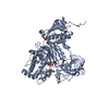
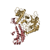
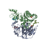
| Entry | Database: PDB / ID: 4yeb | ||||||
|---|---|---|---|---|---|---|---|
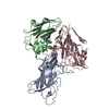
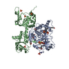
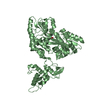
| Title | Structural characterization of a synaptic adhesion complex | ||||||
 Components Components |
| ||||||
 Keywords Keywords |  SIGNALING PROTEIN / Complex / Latrophilin 3 / FLRT3 / Central Nervous System SIGNALING PROTEIN / Complex / Latrophilin 3 / FLRT3 / Central Nervous System | ||||||
| Function / homology |  Function and homology information Function and homology informationproepicardium cell migration involved in pericardium morphogenesis / Downstream signaling of activated FGFR1 / locomotion involved in locomotory behavior / head development / synaptic membrane adhesion / growth cone membrane / maintenance of postsynaptic specialization structure / embryonic morphogenesis / cell-cell adhesion via plasma-membrane adhesion molecules / fibroblast growth factor receptor binding ...proepicardium cell migration involved in pericardium morphogenesis / Downstream signaling of activated FGFR1 / locomotion involved in locomotory behavior / head development / synaptic membrane adhesion / growth cone membrane / maintenance of postsynaptic specialization structure / embryonic morphogenesis / cell-cell adhesion via plasma-membrane adhesion molecules / fibroblast growth factor receptor binding / chemorepellent activity / positive regulation of synapse assembly / neuron projection extension / response to axon injury / fibroblast growth factor receptor signaling pathway / axon terminus / axonal growth cone / synapse assembly / response to cocaine / synaptic membrane / G protein-coupled receptor activity / axon guidance / synapse organization / neuron migration / Schaffer collateral - CA1 synapse / adenylate cyclase-activating G protein-coupled receptor signaling pathway / neuron projection development / cell-cell junction / cell junction / heart development / carbohydrate binding / postsynaptic membrane / postsynaptic density / cell surface receptor signaling pathway / axon / focal adhesion / glutamatergic synapse / calcium ion binding / endoplasmic reticulum membrane / protein homodimerization activity / extracellular space / plasma membrane / cytosolSimilarity search - Function | ||||||
| Biological species |  Mus musculus (house mouse) Mus musculus (house mouse) | ||||||
| Method | X-RAY DIFFRACTION / SYNCHROTRON / MOLECULAR REPLACEMENT / Resolution: 3.19 Å | ||||||
 Authors Authors | Ranaivoson, F.M. / Liu, Q. / Martini, F. / Bergami, F. / von Daake, S. / Li, S. / Lee, D. / Demeler, B. / Hendrickson, W.A. / Comoletti, D. | ||||||
 Citation Citation | Journal: Structure / Year: 2015 Title: Structural and Mechanistic Insights into the Latrophilin3-FLRT3 Complex that Mediates Glutamatergic Synapse Development. Authors: Ranaivoson, F.M. / Liu, Q. / Martini, F. / Bergami, F. / von Daake, S. / Li, S. / Lee, D. / Demeler, B. / Hendrickson, W.A. / Comoletti, D. | ||||||
| History |
|
- Structure visualization
| Structure viewer | Molecule: MolmilJmol/JSmol |
|---|
- Downloads & links
-Download
| PDBx/mmCIF format | 4yeb.cif.gz | 260.8 KB | Display | PDBx/mmCIF format |
|---|---|---|---|---|
| PDB format | pdb4yeb.ent.gz | 212 KB | Display | PDB format |
| PDBx/mmJSON format | 4yeb.json.gz | Tree view | PDBx/mmJSON format | |
| Others |  Other downloads Other downloads |
-Validation report
| Arichive directory | https://data.pdbj.org/pub/pdb/validation_reports/ye/4yebftp://data.pdbj.org/pub/pdb/validation_reports/ye/4yeb | HTTPS FTP |
|---|
-Related structure data
| Related structure data |  4rmkSC  4rmlC 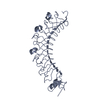 4v2eS S: Starting model for refinement C: citing same article ( |
|---|---|
| Similar structure data |


-Links
 PDBj
PDBj


- Assembly
| Deposited unit | 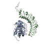
| ||||||||
|---|---|---|---|---|---|---|---|---|---|
| 1 |
| ||||||||
| Unit cell |
|
-Components
| #1: Protein | / Lectomedin-3 Mass: 36632.574 Da / Num. of mol.: 1 Source method: isolated from a genetically manipulated source Source: (gene. exp.) Mus musculus (house mouse) / Gene: Lphn3, Kiaa0768, Lec3 / Cell line (production host): HEK293 / Production host:  Homo sapiens (human) / References: UniProt: Q80TS3 Homo sapiens (human) / References: UniProt: Q80TS3 |
|---|---|
| #2: Protein | Mass: 41677.434 Da / Num. of mol.: 1 Source method: isolated from a genetically manipulated source Source: (gene. exp.) Mus musculus (house mouse) / Gene: Flrt3, mCG_130708 / Cell line (production host): HEK293 / Production host: Homo sapiens (human) / References: UniProt: Q8BGT1 |
| #3: Chemical | ChemComp-CA /   Mass: 40.078 Da / Num. of mol.: 1 / Source method: obtained synthetically / Formula: Ca Mass: 40.078 Da / Num. of mol.: 1 / Source method: obtained synthetically / Formula: Ca |
| #4: Sugar | ChemComp-NAG / N-Acetylglucosamine  Type: D-saccharide, beta linking / Mass: 221.208 Da / Num. of mol.: 1 Type: D-saccharide, beta linking / Mass: 221.208 Da / Num. of mol.: 1Source method: isolated from a genetically manipulated source Formula: C8H15NO6 |
-Experimental details
-Experiment
| Experiment | Method: X-RAY DIFFRACTION |
|---|
- Sample preparation
| Crystal | Density Matthews: 2.3 Å3/Da / Density % sol: 46.55 % |
|---|---|
| Crystal grow | Temperature: 293 K / Method: vapor diffusion, hanging drop / Details: 6 % PEG 3350, 0.2 M NaNO3 |
-Data collection
| Diffraction | Mean temperature: 100 K |
|---|---|
| Diffraction source | Source: SYNCHROTRON / Site: CHESS  / Beamline: F1 / Wavelength: 0.918 Å / Beamline: F1 / Wavelength: 0.918 Å |
| Detector | Type: ADSC QUANTUM 270 / Detector: CCD / Date: Nov 30, 2014 |
| Radiation | Protocol: SINGLE WAVELENGTH / Monochromatic (M) / Laue (L): M / Scattering type: x-ray |
| Radiation wavelength | Wavelength: 0.918 Å / Relative weight: 1 |
| Reflection | Resolution: 3.19→49.33 Å / Num. obs: 12230 / % possible obs: 99.7 % / Redundancy: 1.9 % / Biso Wilson estimate: 138.31 Å2 / Rmerge(I) obs: 0.055 / Net I/σ(I): 9.4 |
| Reflection shell | Resolution: 3.19→3.41 Å / Redundancy: 1.9 % / Rmerge(I) obs: 0.474 / Mean I/σ(I) obs: 1.5 / % possible all: 99 |
- Processing
| Software |
| ||||||||||||||||||||||||||||||||||||||||||||||||||||||||||||||||||||||||||||||||||||||||||||||||||||||||||||||||||||||||||||||||||||||||||||||||||||||||||||||||||||||||||||||||||||||||||||||||||||||||||||||||||||||||||||||||||||||||||||||||||||||||||||||||||||||||||||||||||||||||||||||||||||||||||||||||||||||||||||||||||||||||||||||||||||||||||||||
|---|---|---|---|---|---|---|---|---|---|---|---|---|---|---|---|---|---|---|---|---|---|---|---|---|---|---|---|---|---|---|---|---|---|---|---|---|---|---|---|---|---|---|---|---|---|---|---|---|---|---|---|---|---|---|---|---|---|---|---|---|---|---|---|---|---|---|---|---|---|---|---|---|---|---|---|---|---|---|---|---|---|---|---|---|---|---|---|---|---|---|---|---|---|---|---|---|---|---|---|---|---|---|---|---|---|---|---|---|---|---|---|---|---|---|---|---|---|---|---|---|---|---|---|---|---|---|---|---|---|---|---|---|---|---|---|---|---|---|---|---|---|---|---|---|---|---|---|---|---|---|---|---|---|---|---|---|---|---|---|---|---|---|---|---|---|---|---|---|---|---|---|---|---|---|---|---|---|---|---|---|---|---|---|---|---|---|---|---|---|---|---|---|---|---|---|---|---|---|---|---|---|---|---|---|---|---|---|---|---|---|---|---|---|---|---|---|---|---|---|---|---|---|---|---|---|---|---|---|---|---|---|---|---|---|---|---|---|---|---|---|---|---|---|---|---|---|---|---|---|---|---|---|---|---|---|---|---|---|---|---|---|---|---|---|---|---|---|---|---|---|---|---|---|---|---|---|---|---|---|---|---|---|---|---|---|---|---|---|---|---|---|---|---|---|---|---|---|---|---|---|---|---|---|---|---|---|---|---|---|---|---|---|---|---|---|---|---|---|---|---|---|---|---|---|---|---|---|---|---|---|---|---|---|---|---|---|---|---|---|---|---|---|---|---|---|---|---|---|---|---|---|
| Refinement | Method to determine structure: MOLECULAR REPLACEMENT Starting model: PDB entry 4RMK and 4V2E Resolution: 3.19→14.43 Å / Cor.coef. Fo:Fc: 0.9387 / Cor.coef. Fo:Fc free: 0.8891 / Cross valid method: THROUGHOUT / σ(F): 0 / SU Rfree Blow DPI: 0.653
| ||||||||||||||||||||||||||||||||||||||||||||||||||||||||||||||||||||||||||||||||||||||||||||||||||||||||||||||||||||||||||||||||||||||||||||||||||||||||||||||||||||||||||||||||||||||||||||||||||||||||||||||||||||||||||||||||||||||||||||||||||||||||||||||||||||||||||||||||||||||||||||||||||||||||||||||||||||||||||||||||||||||||||||||||||||||||||||||
| Displacement parameters | Biso mean: 154.44 Å2
| ||||||||||||||||||||||||||||||||||||||||||||||||||||||||||||||||||||||||||||||||||||||||||||||||||||||||||||||||||||||||||||||||||||||||||||||||||||||||||||||||||||||||||||||||||||||||||||||||||||||||||||||||||||||||||||||||||||||||||||||||||||||||||||||||||||||||||||||||||||||||||||||||||||||||||||||||||||||||||||||||||||||||||||||||||||||||||||||
| Refine analyze | Luzzati coordinate error obs: 1.343 Å | ||||||||||||||||||||||||||||||||||||||||||||||||||||||||||||||||||||||||||||||||||||||||||||||||||||||||||||||||||||||||||||||||||||||||||||||||||||||||||||||||||||||||||||||||||||||||||||||||||||||||||||||||||||||||||||||||||||||||||||||||||||||||||||||||||||||||||||||||||||||||||||||||||||||||||||||||||||||||||||||||||||||||||||||||||||||||||||||
| Refinement step | Cycle: LAST / Resolution: 3.19→14.43 Å
| ||||||||||||||||||||||||||||||||||||||||||||||||||||||||||||||||||||||||||||||||||||||||||||||||||||||||||||||||||||||||||||||||||||||||||||||||||||||||||||||||||||||||||||||||||||||||||||||||||||||||||||||||||||||||||||||||||||||||||||||||||||||||||||||||||||||||||||||||||||||||||||||||||||||||||||||||||||||||||||||||||||||||||||||||||||||||||||||
| Refine LS restraints |
| ||||||||||||||||||||||||||||||||||||||||||||||||||||||||||||||||||||||||||||||||||||||||||||||||||||||||||||||||||||||||||||||||||||||||||||||||||||||||||||||||||||||||||||||||||||||||||||||||||||||||||||||||||||||||||||||||||||||||||||||||||||||||||||||||||||||||||||||||||||||||||||||||||||||||||||||||||||||||||||||||||||||||||||||||||||||||||||||
| LS refinement shell | Resolution: 3.19→3.49 Å / Total num. of bins used: 6
| ||||||||||||||||||||||||||||||||||||||||||||||||||||||||||||||||||||||||||||||||||||||||||||||||||||||||||||||||||||||||||||||||||||||||||||||||||||||||||||||||||||||||||||||||||||||||||||||||||||||||||||||||||||||||||||||||||||||||||||||||||||||||||||||||||||||||||||||||||||||||||||||||||||||||||||||||||||||||||||||||||||||||||||||||||||||||||||||
| Refinement TLS params. | Method: refined / Refine-ID: X-RAY DIFFRACTION
| ||||||||||||||||||||||||||||||||||||||||||||||||||||||||||||||||||||||||||||||||||||||||||||||||||||||||||||||||||||||||||||||||||||||||||||||||||||||||||||||||||||||||||||||||||||||||||||||||||||||||||||||||||||||||||||||||||||||||||||||||||||||||||||||||||||||||||||||||||||||||||||||||||||||||||||||||||||||||||||||||||||||||||||||||||||||||||||||
| Refinement TLS group |
|
