 Movie
Movie Controller

[English] 日本語
 Yorodumi
- PDB-4y6x: Structure of Tobacco streak virus coat protein at 2.1 Angstroms r... -
+ Open data

- Basic information
| Entry | Database: PDB / ID: 4y6x | ||||||
|---|---|---|---|---|---|---|---|
| Title | Structure of Tobacco streak virus coat protein at 2.1 Angstroms resolution (C2 crystal form) | ||||||
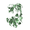
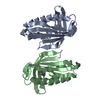
 Components Components | Coat protein | ||||||
 Keywords Keywords |  STRUCTURAL PROTEIN / domain-swapped dimer / jelly roll beta-barrel STRUCTURAL PROTEIN / domain-swapped dimer / jelly roll beta-barrel | ||||||
| Function / homology | Coat protein, Ilarvirus, predicted / Coat protein, Ilarvirus / Ilarvirus coat protein / T=3 icosahedral viral capsid / translational initiation / viral nucleocapsid / ribonucleoprotein complex / RNA binding / Capsid protein Function and homology information Function and homology information | ||||||
| Biological species |  Tobacco streak virus Tobacco streak virus | ||||||
| Method | X-RAY DIFFRACTION / SYNCHROTRON / MOLECULAR REPLACEMENT / Resolution: 2.1 Å | ||||||
 Authors Authors | Gulati, A. / Murthy, M.R.N. | ||||||
| Funding support |  India, 1items India, 1items
| ||||||
 Citation Citation | Journal: J.Struct.Biol. / Year: 2016 Title: Structural studies on tobacco streak virus coat protein: Insights into the pleomorphic nature of ilarviruses Authors: Gulati, A. / Alapati, K. / Murthy, A. / Savithri, H.S. / Murthy, M.R.N. | ||||||
| History |
|
- Structure visualization
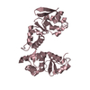
| Structure viewer | Molecule: MolmilJmol/JSmol |
|---|
- Downloads & links
-Download
| PDBx/mmCIF format | 4y6x.cif.gz | 100.7 KB | Display | PDBx/mmCIF format |
|---|---|---|---|---|
| PDB format | pdb4y6x.ent.gz | 75.1 KB | Display | PDB format |
| PDBx/mmJSON format | 4y6x.json.gz | Tree view | PDBx/mmJSON format | |
| Others |  Other downloads Other downloads |
-Validation report
| Arichive directory | https://data.pdbj.org/pub/pdb/validation_reports/y6/4y6xftp://data.pdbj.org/pub/pdb/validation_reports/y6/4y6x | HTTPS FTP |
|---|
-Related structure data
| Related structure data |  4y6tSC S: Starting model for refinement C: citing same article ( |
|---|---|
| Similar structure data |


-Links
 PDBj
PDBj
- Assembly
| Deposited unit | 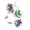
| |||||||||||||||||||||||||||||||||||||||||||||||||||||||||||||||||||||||||||||||||
|---|---|---|---|---|---|---|---|---|---|---|---|---|---|---|---|---|---|---|---|---|---|---|---|---|---|---|---|---|---|---|---|---|---|---|---|---|---|---|---|---|---|---|---|---|---|---|---|---|---|---|---|---|---|---|---|---|---|---|---|---|---|---|---|---|---|---|---|---|---|---|---|---|---|---|---|---|---|---|---|---|---|---|
| 1 | 
| |||||||||||||||||||||||||||||||||||||||||||||||||||||||||||||||||||||||||||||||||
| 2 | 
| |||||||||||||||||||||||||||||||||||||||||||||||||||||||||||||||||||||||||||||||||
| 3 | 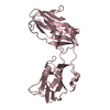
| |||||||||||||||||||||||||||||||||||||||||||||||||||||||||||||||||||||||||||||||||
| Unit cell |
| |||||||||||||||||||||||||||||||||||||||||||||||||||||||||||||||||||||||||||||||||
| Noncrystallographic symmetry (NCS) | NCS domain:
NCS domain segments: Component-ID: 0 / Beg auth comp-ID: SER / Beg label comp-ID: SER / Refine code: 0
NCS ensembles :
|
-Components
| #1: Protein | Mass: 18533.301 Da / Num. of mol.: 3 / Fragment: UNP residues 73-238 Source method: isolated from a genetically manipulated source Source: (gene. exp.) Tobacco streak virus / Production host:  Escherichia coli (E. coli) / References: UniProt: A7UMQ4 Escherichia coli (E. coli) / References: UniProt: A7UMQ4#2: Water | ChemComp-HOH / | Water Mass: 18.015 Da / Num. of mol.: 190 / Source method: isolated from a natural source / Formula: H2O Mass: 18.015 Da / Num. of mol.: 190 / Source method: isolated from a natural source / Formula: H2O |
|---|
-Experimental details
-Experiment
| Experiment | Method: X-RAY DIFFRACTION / Number of used crystals: 1 |
|---|
- Sample preparation
| Crystal | Density Matthews: 2.19 Å3/Da / Density % sol: 43.74 % |
|---|---|
| Crystal grow | Temperature: 295 K / Method: vapor diffusion, hanging drop / pH: 7.5 / Details: 24% PEG 1500, 0.2 M L-Proline, 0.1 M HEPES |
-Data collection
| Diffraction | Mean temperature: 100 K | ||||||||||||||||||||||||||||||||||||||||||||||||||||||||||||||||||||||||||||||||||||||||||||||||||||||||||||||
|---|---|---|---|---|---|---|---|---|---|---|---|---|---|---|---|---|---|---|---|---|---|---|---|---|---|---|---|---|---|---|---|---|---|---|---|---|---|---|---|---|---|---|---|---|---|---|---|---|---|---|---|---|---|---|---|---|---|---|---|---|---|---|---|---|---|---|---|---|---|---|---|---|---|---|---|---|---|---|---|---|---|---|---|---|---|---|---|---|---|---|---|---|---|---|---|---|---|---|---|---|---|---|---|---|---|---|---|---|---|---|---|
| Diffraction source | Source: SYNCHROTRON / Site: ESRF  / Beamline: BM14 / Wavelength: 1.2819 Å / Beamline: BM14 / Wavelength: 1.2819 Å | ||||||||||||||||||||||||||||||||||||||||||||||||||||||||||||||||||||||||||||||||||||||||||||||||||||||||||||||
| Detector | Type: MARMOSAIC 225 mm CCD / Detector: CCD / Date: Sep 28, 2013 | ||||||||||||||||||||||||||||||||||||||||||||||||||||||||||||||||||||||||||||||||||||||||||||||||||||||||||||||
| Radiation | Monochromator: Si(111) monochromator / Protocol: SINGLE WAVELENGTH / Monochromatic (M) / Laue (L): M / Scattering type: x-ray | ||||||||||||||||||||||||||||||||||||||||||||||||||||||||||||||||||||||||||||||||||||||||||||||||||||||||||||||
| Radiation wavelength | Wavelength: 1.2819 Å / Relative weight: 1 | ||||||||||||||||||||||||||||||||||||||||||||||||||||||||||||||||||||||||||||||||||||||||||||||||||||||||||||||
| Reflection | Resolution: 2.1→71.107 Å / Num. all: 27988 / Num. obs: 27988 / % possible obs: 99.8 % / Redundancy: 4.1 % / Rpim(I) all: 0.038 / Rrim(I) all: 0.077 / Rsym value: 0.067 / Net I/av σ(I): 8.822 / Net I/σ(I): 13.1 / Num. measured all: 114040 | ||||||||||||||||||||||||||||||||||||||||||||||||||||||||||||||||||||||||||||||||||||||||||||||||||||||||||||||
| Reflection shell | Diffraction-ID: 1 / Rejects: 0
|
- Processing
| Software |
| |||||||||||||||||||||||||||||||||||||||||||||||||||||||||||||||||||||||||||
|---|---|---|---|---|---|---|---|---|---|---|---|---|---|---|---|---|---|---|---|---|---|---|---|---|---|---|---|---|---|---|---|---|---|---|---|---|---|---|---|---|---|---|---|---|---|---|---|---|---|---|---|---|---|---|---|---|---|---|---|---|---|---|---|---|---|---|---|---|---|---|---|---|---|---|---|---|
| Refinement | Method to determine structure: MOLECULAR REPLACEMENT Starting model: 4Y6T Resolution: 2.1→28.93 Å / Cor.coef. Fo:Fc: 0.961 / Cor.coef. Fo:Fc free: 0.942 / WRfactor Rfree: 0.225 / WRfactor Rwork: 0.1755 / FOM work R set: 0.851 / SU B: 4.845 / SU ML: 0.128 / SU R Cruickshank DPI: 0.1981 / SU Rfree: 0.177 / Cross valid method: THROUGHOUT / σ(F): 0 / ESU R: 0.198 / ESU R Free: 0.177 / Stereochemistry target values: MAXIMUM LIKELIHOOD Details: HYDROGENS HAVE BEEN ADDED IN THE RIDING POSITIONS U VALUES : REFINED INDIVIDUALLY
| |||||||||||||||||||||||||||||||||||||||||||||||||||||||||||||||||||||||||||
| Solvent computation | Ion probe radii: 0.8 Å / Shrinkage radii: 0.8 Å / VDW probe radii: 1.2 Å / Solvent model: MASK | |||||||||||||||||||||||||||||||||||||||||||||||||||||||||||||||||||||||||||
| Displacement parameters | Biso max: 100.64 Å2 / Biso mean: 39.856 Å2 / Biso min: 17.52 Å2
| |||||||||||||||||||||||||||||||||||||||||||||||||||||||||||||||||||||||||||
| Refinement step | Cycle: final / Resolution: 2.1→28.93 Å
| |||||||||||||||||||||||||||||||||||||||||||||||||||||||||||||||||||||||||||
| Refine LS restraints |
| |||||||||||||||||||||||||||||||||||||||||||||||||||||||||||||||||||||||||||
| Refine LS restraints NCS | Refine-ID: X-RAY DIFFRACTION / Type: interatomic distance / Weight position: 0.05
| |||||||||||||||||||||||||||||||||||||||||||||||||||||||||||||||||||||||||||
| LS refinement shell | Resolution: 2.1→2.155 Å / Total num. of bins used: 20
|
