 Movie
Movie Controller

[English] 日本語
 Yorodumi
- PDB-4y6c: Q17M crystal structure of Podosopora anserina putative kinesin li... -
+ Open data

- Basic information
| Entry | Database: PDB / ID: 4y6c | ||||||
|---|---|---|---|---|---|---|---|
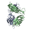
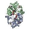
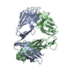
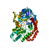
| Title | Q17M crystal structure of Podosopora anserina putative kinesin light chain nearly identical TPR-like repeats | ||||||
 Components Components | ANSERINA PUTATIVE KINESIN LIGHT chain | ||||||
 Keywords Keywords | UNKNOWN FUNCTION /  Tetratricopeptide repeat / 42PR / TPR Tetratricopeptide repeat / 42PR / TPR | ||||||
| Function / homology |  Function and homology information Function and homology information | ||||||
| Biological species |  Podospora anserina (fungus) Podospora anserina (fungus) | ||||||
| Method | X-RAY DIFFRACTION / SYNCHROTRON / SAD / Resolution: 1.772 Å | ||||||
 Authors Authors | Marold, J.D. / Kavran, J.M. / Bowman, G.D. / Barrick, D. | ||||||
 Citation Citation | Journal: Structure / Year: 2015 Title: A Naturally Occurring Repeat Protein with High Internal Sequence Identity Defines a New Class of TPR-like Proteins. Authors: Marold, J.D. / Kavran, J.M. / Bowman, G.D. / Barrick, D. | ||||||
| History |
|
- Structure visualization
| Structure viewer | Molecule: MolmilJmol/JSmol |
|---|
- Downloads & links
-Download
| PDBx/mmCIF format | 4y6c.cif.gz | 101 KB | Display | PDBx/mmCIF format |
|---|---|---|---|---|
| PDB format | pdb4y6c.ent.gz | 81.8 KB | Display | PDB format |
| PDBx/mmJSON format | 4y6c.json.gz | Tree view | PDBx/mmJSON format | |
| Others |  Other downloads Other downloads |
-Validation report
| Arichive directory | https://data.pdbj.org/pub/pdb/validation_reports/y6/4y6cftp://data.pdbj.org/pub/pdb/validation_reports/y6/4y6c | HTTPS FTP |
|---|
-Related structure data


-Links
 PDBj
PDBj


- Assembly
| Deposited unit | 
| ||||||||
|---|---|---|---|---|---|---|---|---|---|
| 1 | 
| ||||||||
| Unit cell |
| ||||||||
| Details | determined by Analytical Ultracentrifugation Sedimentation Velocity |
-Components
| #1: Protein | Mass: 26023.572 Da / Num. of mol.: 1 Source method: isolated from a genetically manipulated source Source: (gene. exp.) Podospora anserina (fungus) / Strain: S mat+ / Gene: CDP31375.1 / Plasmid: pET15b / Cell line (production host): ROSETTA 2 / Production host:  Escherichia coli (E. coli) / Strain (production host): DE3 / References: UniProt: A0A090CRQ5*PLUS Escherichia coli (E. coli) / Strain (production host): DE3 / References: UniProt: A0A090CRQ5*PLUS | ||
|---|---|---|---|
| #2: Chemical | Sulfate  Mass: 96.063 Da / Num. of mol.: 2 / Source method: obtained synthetically / Formula: SO4 Mass: 96.063 Da / Num. of mol.: 2 / Source method: obtained synthetically / Formula: SO4#3: Water | ChemComp-HOH / | Water Mass: 18.015 Da / Num. of mol.: 169 / Source method: isolated from a natural source / Formula: H2O Mass: 18.015 Da / Num. of mol.: 169 / Source method: isolated from a natural source / Formula: H2O |
-Experimental details
-Experiment
| Experiment | Method: X-RAY DIFFRACTION |
|---|
- Sample preparation
| Crystal | Density Matthews: 2.21 Å3/Da / Density % sol: 44.39 % |
|---|---|
| Crystal grow | Temperature: 295.16 K / Method: vapor diffusion, hanging drop / pH: 6.5 / Details: 0.1 M MES, 35% (w/v) PEG 4000 |
-Data collection
| Diffraction | Mean temperature: 80 K |
|---|---|
| Diffraction source | Source: SYNCHROTRON / Site: NSLS  / Beamline: X29A / Wavelength: 0.978 Å / Beamline: X29A / Wavelength: 0.978 Å |
| Detector | Type: ADSC QUANTUM 315r / Detector: CCD / Date: Apr 30, 2013 |
| Radiation | Monochromator: Rosenbaum-Rock double crystal sagittal / Protocol: SINGLE WAVELENGTH / Monochromatic (M) / Laue (L): M / Scattering type: x-ray |
| Radiation wavelength | Wavelength: 0.978 Å / Relative weight: 1 |
| Reflection | Resolution: 1.772→39.76 Å / Num. all: 23011 / Num. obs: 23013 / % possible obs: 99.97 % / Redundancy: 14.3 % / Rmerge(I) obs: 0.175 / Rsym value: 0.172 / Net I/σ(I): 17.7 |
| Reflection shell | Resolution: 1.773→1.836 Å / Redundancy: 13.9 % / Rmerge(I) obs: 0.348 / Mean I/σ(I) obs: 11.4 / % possible all: 99.48 |
- Processing
| Software |
| |||||||||||||||||||||||||||||||||||||||||||||||||||||||||||||||
|---|---|---|---|---|---|---|---|---|---|---|---|---|---|---|---|---|---|---|---|---|---|---|---|---|---|---|---|---|---|---|---|---|---|---|---|---|---|---|---|---|---|---|---|---|---|---|---|---|---|---|---|---|---|---|---|---|---|---|---|---|---|---|---|---|
| Refinement | Method to determine structure: SAD / Resolution: 1.772→39.76 Å / SU ML: 0.17 / Cross valid method: FREE R-VALUE / σ(F): 1.39 / Phase error: 18.04 / Stereochemistry target values: ML
| |||||||||||||||||||||||||||||||||||||||||||||||||||||||||||||||
| Solvent computation | Shrinkage radii: 0.9 Å / VDW probe radii: 1.11 Å / Solvent model: FLAT BULK SOLVENT MODEL | |||||||||||||||||||||||||||||||||||||||||||||||||||||||||||||||
| Refinement step | Cycle: LAST / Resolution: 1.772→39.76 Å
| |||||||||||||||||||||||||||||||||||||||||||||||||||||||||||||||
| Refine LS restraints |
| |||||||||||||||||||||||||||||||||||||||||||||||||||||||||||||||
| LS refinement shell |
| |||||||||||||||||||||||||||||||||||||||||||||||||||||||||||||||
| Refinement TLS params. | Method: refined / Origin x: -10.48 Å / Origin y: -40.3489 Å / Origin z: -0.4465 Å
| |||||||||||||||||||||||||||||||||||||||||||||||||||||||||||||||
| Refinement TLS group | Selection details: all |
