 Movie
Movie Controller


+ Open data

- Basic information
| Entry | Database: PDB / ID: 4y66 | ||||||
|---|---|---|---|---|---|---|---|
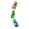
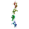
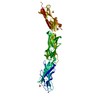
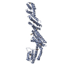
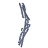
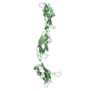
| Title | Crystal structure of Giardia lamblia Hop2-Mnd1 complex | ||||||
 Components Components |
| ||||||
 Keywords Keywords |  CELL CYCLE CELL CYCLE | ||||||
| Function / homology |  Function and homology information Function and homology informationreciprocal meiotic recombination / double-stranded DNA binding / nucleusSimilarity search - Function | ||||||
| Biological species |  Giardia lamblia ATCC 50803 (eukaryote) Giardia lamblia ATCC 50803 (eukaryote)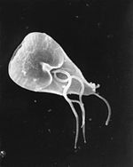 Giardia lamblia (eukaryote) Giardia lamblia (eukaryote) | ||||||
| Method | X-RAY DIFFRACTION / SYNCHROTRON / SAD / Resolution: 3.2 Å | ||||||
 Authors Authors | Kang, H.A. / Shin, H.C. / Oh, B.H. | ||||||
 Citation Citation | Journal: Nucleic Acids Res. / Year: 2015 Title: Crystal structure of Hop2-Mnd1 and mechanistic insights into its role in meiotic recombination Authors: Kang, H.A. / Shin, H.C. / Kalantzi, A.S. / Toseland, C.P. / Kim, H.M. / Gruber, S. / Peraro, M.D. / Oh, B.H. | ||||||
| History |
|
- Structure visualization
| Structure viewer | Molecule: MolmilJmol/JSmol |
|---|
- Downloads & links
-Download
| PDBx/mmCIF format | 4y66.cif.gz | 371.3 KB | Display | PDBx/mmCIF format |
|---|---|---|---|---|
| PDB format | pdb4y66.ent.gz | 301.9 KB | Display | PDB format |
| PDBx/mmJSON format | 4y66.json.gz | Tree view | PDBx/mmJSON format | |
| Others |  Other downloads Other downloads |
-Validation report
| Arichive directory | https://data.pdbj.org/pub/pdb/validation_reports/y6/4y66ftp://data.pdbj.org/pub/pdb/validation_reports/y6/4y66 | HTTPS FTP |
|---|
-Related structure data
| Similar structure data |
|---|


-Links
 PDBj
PDBj- Assembly
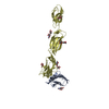
| Deposited unit | 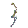
| ||||||||
|---|---|---|---|---|---|---|---|---|---|
| 1 | 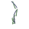
| ||||||||
| 2 | 
| ||||||||
| 3 | 
| ||||||||
| Unit cell |
|
-Components
| #1: Protein | Mass: 23311.662 Da / Num. of mol.: 3 Source method: isolated from a genetically manipulated source Source: (gene. exp.) Giardia lamblia ATCC 50803 (eukaryote) / Strain: ATCC 50803 / Gene: GL50803_6626 / Production host:  Escherichia coli (E. coli) / References: UniProt: E2RTU1 Escherichia coli (E. coli) / References: UniProt: E2RTU1#2: Protein | Mass: 25739.529 Da / Num. of mol.: 3 Source method: isolated from a genetically manipulated source Source: (gene. exp.) Giardia lamblia (eukaryote) / Strain: ATCC 50803 / Gene: GSB_17044 / Production host: Escherichia coli (E. coli) / References: UniProt: V6TR15 |
|---|
-Experimental details
-Experiment
| Experiment | Method: X-RAY DIFFRACTION |
|---|
- Sample preparation
| Crystal | Density Matthews: 4 Å3/Da / Density % sol: 69.22 % |
|---|---|
| Crystal grow | Temperature: 291 K / Method: vapor diffusion, hanging drop / Details: PEG 3350, Tascimate, Tris |
-Data collection
| Diffraction | Mean temperature: 110 K |
|---|---|
| Diffraction source | Source: SYNCHROTRON / Site: PAL/PLS  / Beamline: 5C (4A) / Wavelength: 0.97889 Å / Beamline: 5C (4A) / Wavelength: 0.97889 Å |
| Detector | Type: ADSC QUANTUM 210 / Detector: CCD / Date: Jul 10, 2013 |
| Radiation | Protocol: SINGLE WAVELENGTH / Monochromatic (M) / Laue (L): M / Scattering type: x-ray |
| Radiation wavelength | Wavelength: 0.97889 Å / Relative weight: 1 |
| Reflection | Resolution: 3.2→50 Å / Num. obs: 34750 / % possible obs: 87 % / Redundancy: 3.8 % / Net I/σ(I): 22.1 |
| Reflection shell | Highest resolution: 3.2 Å / Redundancy: 1.9 % / Mean I/σ(I) obs: 3.1 / Rsym value: 0.272 / % possible all: 64.9 |
- Processing
| Software |
| |||||||||||||||||||||||||||||||||||||||||||||||||||||||||||||||||||||||||||||||||||||||||||
|---|---|---|---|---|---|---|---|---|---|---|---|---|---|---|---|---|---|---|---|---|---|---|---|---|---|---|---|---|---|---|---|---|---|---|---|---|---|---|---|---|---|---|---|---|---|---|---|---|---|---|---|---|---|---|---|---|---|---|---|---|---|---|---|---|---|---|---|---|---|---|---|---|---|---|---|---|---|---|---|---|---|---|---|---|---|---|---|---|---|---|---|---|
| Refinement | Method to determine structure: SAD / Resolution: 3.2→40.65 Å / SU ML: 0.52 / Cross valid method: FREE R-VALUE / σ(F): 1.78 / Phase error: 29.46 / Stereochemistry target values: ML
| |||||||||||||||||||||||||||||||||||||||||||||||||||||||||||||||||||||||||||||||||||||||||||
| Solvent computation | Shrinkage radii: 0.9 Å / VDW probe radii: 1.11 Å / Solvent model: FLAT BULK SOLVENT MODEL | |||||||||||||||||||||||||||||||||||||||||||||||||||||||||||||||||||||||||||||||||||||||||||
| Refinement step | Cycle: LAST / Resolution: 3.2→40.65 Å
| |||||||||||||||||||||||||||||||||||||||||||||||||||||||||||||||||||||||||||||||||||||||||||
| Refine LS restraints |
| |||||||||||||||||||||||||||||||||||||||||||||||||||||||||||||||||||||||||||||||||||||||||||
| LS refinement shell |
| |||||||||||||||||||||||||||||||||||||||||||||||||||||||||||||||||||||||||||||||||||||||||||
| Refinement TLS params. | Method: refined / Origin x: 20.6299 Å / Origin y: 56.7855 Å / Origin z: -50.0752 Å
| |||||||||||||||||||||||||||||||||||||||||||||||||||||||||||||||||||||||||||||||||||||||||||
| Refinement TLS group | Selection details: ALL |
