 Movie
Movie Controller


+ Open data

- Basic information
| Entry | Database: PDB / ID: 4y3u | ||||||
|---|---|---|---|---|---|---|---|
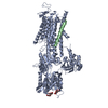
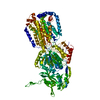
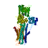
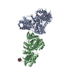
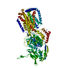
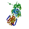
| Title | The structure of phospholamban bound to the calcium pump SERCA1a | ||||||
 Components Components |
| ||||||
 Keywords Keywords |  MEMBRANE PROTEIN / Ca-ATPase / SERCA1a MEMBRANE PROTEIN / Ca-ATPase / SERCA1a | ||||||
| Function / homology |  Function and homology information Function and homology informationIon homeostasis / Ion transport by P-type ATPases / negative regulation of calcium ion import into sarcoplasmic reticulum / negative regulation of ATPase-coupled calcium transmembrane transporter activity / adenylate cyclase-activating adrenergic receptor signaling pathway involved in heart process / regulation of relaxation of muscle / regulation of the force of heart contraction by cardiac conduction / calcium ion-transporting ATPase complex / acrosome assembly / negative regulation of calcium ion transmembrane transporter activity ...Ion homeostasis / Ion transport by P-type ATPases / negative regulation of calcium ion import into sarcoplasmic reticulum / negative regulation of ATPase-coupled calcium transmembrane transporter activity / adenylate cyclase-activating adrenergic receptor signaling pathway involved in heart process / regulation of relaxation of muscle / regulation of the force of heart contraction by cardiac conduction / calcium ion-transporting ATPase complex / acrosome assembly / negative regulation of calcium ion transmembrane transporter activity / positive regulation of cardiac muscle cell contraction / positive regulation of calcium ion import into sarcoplasmic reticulum / H zone / positive regulation of fast-twitch skeletal muscle fiber contraction / calcium ion import into sarcoplasmic reticulum / negative regulation of striated muscle contraction / regulation of striated muscle contraction / positive regulation of ATPase-coupled calcium transmembrane transporter activity / regulation of calcium ion import / P-type Ca2+ transporter / P-type calcium transporter activity / ATPase inhibitor activity / negative regulation of ATP-dependent activity / regulation of cardiac muscle cell contraction / cardiac muscle tissue development / I band / negative regulation of heart rate / muscle cell cellular homeostasis / regulation of calcium ion transport / endoplasmic reticulum-Golgi intermediate compartment / regulation of cardiac muscle contraction by regulation of the release of sequestered calcium ion / Notch signaling pathway / sarcoplasmic reticulum membrane / sarcoplasmic reticulum / mitochondrial membrane / intracellular calcium ion homeostasis / calcium ion transport / calcium ion binding / endoplasmic reticulum membrane / perinuclear region of cytoplasm / endoplasmic reticulum / ATP hydrolysis activity / protein homodimerization activity / ATP binding / membraneSimilarity search - Function | ||||||
| Biological species |  Canis familiaris (dog)Oryctolagus cuniculus (rabbit) Canis familiaris (dog)Oryctolagus cuniculus (rabbit) | ||||||
| Method | X-RAY DIFFRACTION / SYNCHROTRON / FOURIER SYNTHESIS / Resolution: 3.51 Å | ||||||
 Authors Authors | Hurley, T.D. | ||||||
| Funding support |  United States, 1items United States, 1items
| ||||||
 Citation Citation | Journal: J. Biol. Chem. / Year: 2013 Title: The structural basis for phospholamban inhibition of the calcium pump in sarcoplasmic reticulum. Authors: Akin, B.L. / Hurley, T.D. / Chen, Z. / Jones, L.R. | ||||||
| History |
|
- Structure visualization
| Structure viewer | Molecule: MolmilJmol/JSmol |
|---|
- Downloads & links
-Download
| PDBx/mmCIF format | 4y3u.cif.gz | 403.4 KB | Display | PDBx/mmCIF format |
|---|---|---|---|---|
| PDB format | pdb4y3u.ent.gz | 335.4 KB | Display | PDB format |
| PDBx/mmJSON format | 4y3u.json.gz | Tree view | PDBx/mmJSON format | |
| Others |  Other downloads Other downloads |
-Validation report
| Arichive directory | https://data.pdbj.org/pub/pdb/validation_reports/y3/4y3uftp://data.pdbj.org/pub/pdb/validation_reports/y3/4y3u | HTTPS FTP |
|---|
-Related structure data
| Related structure data |  4kytSC S: Starting model for refinement C: citing same article ( |
|---|---|
| Similar structure data |


-Links
 PDBj
PDBj


- Assembly
| Deposited unit | 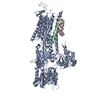
| ||||||||
|---|---|---|---|---|---|---|---|---|---|
| 1 |
| ||||||||
| Unit cell |
|
-Components
| #1: Protein | Mass: 109602.578 Da / Num. of mol.: 1 / Source method: isolated from a natural source / Source: (natural) Oryctolagus cuniculus (rabbit) / Organ: MuscleSkeletal muscle / Tissue: skeletal muscle / References: UniProt: P04191, EC: 3.6.3.8 |
|---|---|
| #2: Protein/peptide | Mass: 5859.158 Da / Num. of mol.: 1 Source method: isolated from a genetically manipulated source Source: (gene. exp.) Canis familiaris (dog) / Gene: PLN / Plasmid: PVL1393 / Cell line (production host): SF21 / Production host:   Spodoptera frugiperda (fall armyworm) / References: UniProt: P61012 Spodoptera frugiperda (fall armyworm) / References: UniProt: P61012 |
| #3: Protein/peptide | Mass: 1975.426 Da / Num. of mol.: 1 Source method: isolated from a genetically manipulated source Details: This is a second copy of phospholamban bound in this structure, but the side chain electron density is insufficient to assign the correct sequence register Source: (gene. exp.) Canis familiaris (dog) / Gene: PLN / Production host: Spodoptera frugiperda (fall armyworm) |
| #4: Chemical | ChemComp-K /   Mass: 39.098 Da / Num. of mol.: 1 / Source method: obtained synthetically / Formula: K Mass: 39.098 Da / Num. of mol.: 1 / Source method: obtained synthetically / Formula: K |
-Experimental details
-Experiment
| Experiment | Method: X-RAY DIFFRACTION |
|---|
- Sample preparation
| Crystal | Density Matthews: 3.73 Å3/Da / Density % sol: 67.1 % |
|---|---|
| Crystal grow | Temperature: 277 K / Method: vapor diffusion, hanging drop / pH: 7 Details: 1 UL OF SERCA1A AT 15 MG/ML IN 2% N- NONYL-BETA-D-MALTOPYRANOSIDE (NONYL MALTOSIDE) (ANATRACE), 20% GLYCEROL, 100 MM MOPS (PH 7.0), 0.12 M SUCROSE, 80 MM KCL, 3 MM MGCL2, AND 2.8 MM EGTA WAS ...Details: 1 UL OF SERCA1A AT 15 MG/ML IN 2% N- NONYL-BETA-D-MALTOPYRANOSIDE (NONYL MALTOSIDE) (ANATRACE), 20% GLYCEROL, 100 MM MOPS (PH 7.0), 0.12 M SUCROSE, 80 MM KCL, 3 MM MGCL2, AND 2.8 MM EGTA WAS MIXED WITH 1 UL OF PHOSPHOLAMBAN AT 2.1 MG/ML IN 20 MM MOPS (PH 7.2), 20% GLYCEROL, AND 0.1 % DECYLMALTOSIDE OR 0.01% DODECYL MALTOSIDE. THIS PROTEIN MIXTURE WAS THEN ADDED TO AN EQUAL VOLUME OF CRYSTALLIZATION LIQUOR; 15 % GLYCEROL, 17% (W/V) PEG-2000, 200MM NAOAC, AND 5 MM BETA-MERCOPTOETHANOL) |
-Data collection
| Diffraction | Mean temperature: 100 K |
|---|---|
| Diffraction source | Source: SYNCHROTRON / Site: APS / Beamline: 19-ID / Wavelength: 0.987 Å |
| Detector | Type: ADSC QUANTUM 315r / Detector: CCD / Date: Aug 10, 2013 |
| Radiation | Protocol: SINGLE WAVELENGTH / Monochromatic (M) / Laue (L): M / Scattering type: x-ray |
| Radiation wavelength | Wavelength: 0.987 Å / Relative weight: 1 |
| Reflection | Resolution: 3.5→50 Å / Num. obs: 23282 / % possible obs: 99 % / Observed criterion σ(F): 0.2 / Observed criterion σ(I): 0.2 / Redundancy: 4.7 % / Rmerge(I) obs: 0.087 / Net I/σ(I): 16.46 |
| Reflection shell | Resolution: 3.5→3.56 Å / Redundancy: 4.2 % / Rmerge(I) obs: 0.543 / Mean I/σ(I) obs: 2.47 / % possible all: 100 |
- Processing
| Software |
| ||||||||||||||||||||||||||||||||||||||||||||||||||||||||||||||||||||||||||||||||||||||||||||||||||||||||||||||||||||||||||||||||||||||||||||||||||||||||||||||||||||||||||||||||||||||
|---|---|---|---|---|---|---|---|---|---|---|---|---|---|---|---|---|---|---|---|---|---|---|---|---|---|---|---|---|---|---|---|---|---|---|---|---|---|---|---|---|---|---|---|---|---|---|---|---|---|---|---|---|---|---|---|---|---|---|---|---|---|---|---|---|---|---|---|---|---|---|---|---|---|---|---|---|---|---|---|---|---|---|---|---|---|---|---|---|---|---|---|---|---|---|---|---|---|---|---|---|---|---|---|---|---|---|---|---|---|---|---|---|---|---|---|---|---|---|---|---|---|---|---|---|---|---|---|---|---|---|---|---|---|---|---|---|---|---|---|---|---|---|---|---|---|---|---|---|---|---|---|---|---|---|---|---|---|---|---|---|---|---|---|---|---|---|---|---|---|---|---|---|---|---|---|---|---|---|---|---|---|---|---|
| Refinement | Method to determine structure: FOURIER SYNTHESIS Starting model: PDB entry 4KYT Resolution: 3.51→48.79 Å / Cor.coef. Fo:Fc: 0.894 / Cor.coef. Fo:Fc free: 0.852 / SU B: 56.647 / SU ML: 0.404 / Cross valid method: THROUGHOUT / ESU R Free: 0.592 / Stereochemistry target values: MAXIMUM LIKELIHOOD
| ||||||||||||||||||||||||||||||||||||||||||||||||||||||||||||||||||||||||||||||||||||||||||||||||||||||||||||||||||||||||||||||||||||||||||||||||||||||||||||||||||||||||||||||||||||||
| Solvent computation | Ion probe radii: 0.8 Å / Shrinkage radii: 0.8 Å / VDW probe radii: 1.2 Å / Solvent model: MASK | ||||||||||||||||||||||||||||||||||||||||||||||||||||||||||||||||||||||||||||||||||||||||||||||||||||||||||||||||||||||||||||||||||||||||||||||||||||||||||||||||||||||||||||||||||||||
| Displacement parameters | Biso mean: 135.931 Å2
| ||||||||||||||||||||||||||||||||||||||||||||||||||||||||||||||||||||||||||||||||||||||||||||||||||||||||||||||||||||||||||||||||||||||||||||||||||||||||||||||||||||||||||||||||||||||
| Refinement step | Cycle: 1 / Resolution: 3.51→48.79 Å
| ||||||||||||||||||||||||||||||||||||||||||||||||||||||||||||||||||||||||||||||||||||||||||||||||||||||||||||||||||||||||||||||||||||||||||||||||||||||||||||||||||||||||||||||||||||||
| Refine LS restraints |
|