 Movie
Movie Controller

[English] 日本語
 Yorodumi
- PDB-4y32: Crystal structure of C-terminal modified Tau peptide-hybrid 109B ... -
+ Open data

- Basic information
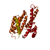
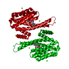
| Entry | Database: PDB / ID: 4y32 | |||||||||
|---|---|---|---|---|---|---|---|---|---|---|
| Title | Crystal structure of C-terminal modified Tau peptide-hybrid 109B with 14-3-3sigma | |||||||||
 Components Components |
| |||||||||
 Keywords Keywords |  SIGNALING PROTEIN / Peptide-hybrid / Inhibitor / Protein-protein interaction / Peptide binding protein / Adapter protein / 14-3-3 SIGNALING PROTEIN / Peptide-hybrid / Inhibitor / Protein-protein interaction / Peptide binding protein / Adapter protein / 14-3-3 | |||||||||
| Function / homology |  Function and homology information Function and homology informationplus-end-directed organelle transport along microtubule / axonal transport / histone-dependent DNA binding / neurofibrillary tangle assembly / positive regulation of diacylglycerol kinase activity / negative regulation of establishment of protein localization to mitochondrion / neurofibrillary tangle / positive regulation of protein localization to synapse / microtubule lateral binding / tubulin complex ...plus-end-directed organelle transport along microtubule / axonal transport / histone-dependent DNA binding / neurofibrillary tangle assembly / positive regulation of diacylglycerol kinase activity / negative regulation of establishment of protein localization to mitochondrion / neurofibrillary tangle / positive regulation of protein localization to synapse / microtubule lateral binding / tubulin complex / phosphatidylinositol bisphosphate binding / main axon / regulation of long-term synaptic depression / negative regulation of kinase activity / negative regulation of tubulin deacetylation / generation of neurons / regulation of chromosome organization / positive regulation of protein localization / rRNA metabolic process / internal protein amino acid acetylation / regulation of mitochondrial fission / intracellular distribution of mitochondria / axonal transport of mitochondrion / axon development / regulation of epidermal cell division / protein kinase C inhibitor activity / central nervous system neuron development / positive regulation of epidermal cell differentiation / keratinocyte development / keratinization / regulation of microtubule polymerization / microtubule polymerization / minor groove of adenine-thymine-rich DNA binding / lipoprotein particle binding / dynactin binding / glial cell projection / negative regulation of mitochondrial membrane potential / apolipoprotein binding / Regulation of localization of FOXO transcription factors / keratinocyte proliferation / protein polymerization / negative regulation of mitochondrial fission / axolemma / phosphoserine residue binding / Activation of BAD and translocation to mitochondria / negative regulation of keratinocyte proliferation / Caspase-mediated cleavage of cytoskeletal proteins / regulation of microtubule polymerization or depolymerization / establishment of skin barrier / positive regulation of axon extension / SARS-CoV-2 targets host intracellular signalling and regulatory pathways / Chk1/Chk2(Cds1) mediated inactivation of Cyclin B:Cdk1 complex / regulation of microtubule cytoskeleton organization / supramolecular fiber organization / Activation of AMPK downstream of NMDARs / stress granule assembly / cytoplasmic microtubule organization / regulation of cellular response to heat / protein kinase A signaling / negative regulation of stem cell proliferation / SARS-CoV-1 targets host intracellular signalling and regulatory pathways / axon cytoplasm / RHO GTPases activate PKNs / regulation of calcium-mediated signaling / positive regulation of microtubule polymerization / cellular response to brain-derived neurotrophic factor stimulus / somatodendritic compartment / synapse assembly / protein export from nucleus / negative regulation of innate immune response / phosphatidylinositol binding / nuclear periphery / protein sequestering activity / TP53 Regulates Transcription of Genes Involved in G2 Cell Cycle Arrest / cellular response to nerve growth factor stimulus / release of cytochrome c from mitochondria / positive regulation of superoxide anion generation / positive regulation of protein export from nucleus / protein phosphatase 2A binding / stem cell proliferation / regulation of autophagy / Translocation of SLC2A4 (GLUT4) to the plasma membrane / astrocyte activation / TP53 Regulates Metabolic Genes / response to lead ion / synapse organization / microglial cell activation / negative regulation of protein kinase activity / Hsp90 protein binding / regulation of synaptic plasticity / PKR-mediated signaling / negative regulation of cysteine-type endopeptidase activity involved in apoptotic process / protein homooligomerization / cytoplasmic ribonucleoprotein granule / memory / microtubule cytoskeleton organization / cellular response to reactive oxygen species / SH3 domain binding / activation of cysteine-type endopeptidase activity involved in apoptotic process / neuron projection developmentSimilarity search - Function | |||||||||
| Biological species |  Homo sapiens (human) Homo sapiens (human) | |||||||||
| Method | X-RAY DIFFRACTION / SYNCHROTRON / MOLECULAR REPLACEMENT / molecular replacement / Resolution: 1.7 Å | |||||||||
 Authors Authors | Bartel, M. / Milroy, L. / Bier, D. / Brunsveld, L. / Ottmann, C. | |||||||||
 Citation Citation | Journal: Angew.Chem.Int.Ed.Engl. / Year: 2015 Title: Stabilizer-Guided Inhibition of Protein-Protein Interactions. Authors: Milroy, L.G. / Bartel, M. / Henen, M.A. / Leysen, S. / Adriaans, J.M. / Brunsveld, L. / Landrieu, I. / Ottmann, C. | |||||||||
| History |
|
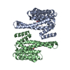
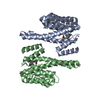
- Structure visualization
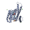
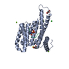
| Structure viewer | Molecule: MolmilJmol/JSmol |
|---|
- Downloads & links
-Download
| PDBx/mmCIF format | 4y32.cif.gz | 131.1 KB | Display | PDBx/mmCIF format |
|---|---|---|---|---|
| PDB format | pdb4y32.ent.gz | 100 KB | Display | PDB format |
| PDBx/mmJSON format | 4y32.json.gz | Tree view | PDBx/mmJSON format | |
| Others |  Other downloads Other downloads |
-Validation report
| Arichive directory | https://data.pdbj.org/pub/pdb/validation_reports/y3/4y32ftp://data.pdbj.org/pub/pdb/validation_reports/y3/4y32 | HTTPS FTP |
|---|
-Related structure data
| Related structure data | 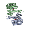 4y3bC 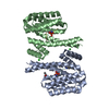 4y5iC 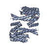 5hf3C 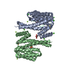 4fl5S  4y3v S: Starting model for refinement C: citing same article ( |
|---|---|
| Similar structure data |


-Links
 PDBj
PDBj


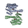
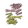
- Assembly
| Deposited unit | 
| ||||||||
|---|---|---|---|---|---|---|---|---|---|
| 1 | 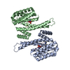
| ||||||||
| Unit cell |
|
-Components
| #1: Protein | Mass: 26542.914 Da / Num. of mol.: 2 Source method: isolated from a genetically manipulated source Source: (gene. exp.) Homo sapiens (human) / Gene: SFN, HME1 / Production host:  Escherichia coli (E. coli) / References: UniProt: P31947 Escherichia coli (E. coli) / References: UniProt: P31947#2: Protein/peptide | Mass: 851.861 Da / Num. of mol.: 2 / Source method: obtained synthetically Details: The C-terminal proline contains chemical modifications Source: (synth.) Homo sapiens (human) / References: UniProt: P10636*PLUS#3: Chemical | ChemComp-49F / ( |   Mass: 129.200 Da / Num. of mol.: 1 / Source method: obtained synthetically / Formula: C7H15NO Mass: 129.200 Da / Num. of mol.: 1 / Source method: obtained synthetically / Formula: C7H15NO#4: Water | ChemComp-HOH / | Water Mass: 18.015 Da / Num. of mol.: 761 / Source method: isolated from a natural source / Formula: H2O Mass: 18.015 Da / Num. of mol.: 761 / Source method: isolated from a natural source / Formula: H2O |
|---|
-Experimental details
-Experiment
| Experiment | Method: X-RAY DIFFRACTION / Number of used crystals: 1 |
|---|
- Sample preparation
| Crystal | Density Matthews: 2.62 Å3/Da / Density % sol: 52.97 % |
|---|---|
| Crystal grow | Temperature: 277 K / Method: vapor diffusion, hanging drop / pH: 7.1 Details: 0.1 M Hepes/NaOH pH 7.1, 0.19 M CaCl2, 24% PEG 400, 5% glycerol |
-Data collection
| Diffraction | Mean temperature: 100 K | ||||||||||||||||||||||||||||||||||||||||||||||||||||||||||||||||||||||||||||||||||||||||||||||||||||||||||||||||||||||||||||||||||||||||||||||||||||||
|---|---|---|---|---|---|---|---|---|---|---|---|---|---|---|---|---|---|---|---|---|---|---|---|---|---|---|---|---|---|---|---|---|---|---|---|---|---|---|---|---|---|---|---|---|---|---|---|---|---|---|---|---|---|---|---|---|---|---|---|---|---|---|---|---|---|---|---|---|---|---|---|---|---|---|---|---|---|---|---|---|---|---|---|---|---|---|---|---|---|---|---|---|---|---|---|---|---|---|---|---|---|---|---|---|---|---|---|---|---|---|---|---|---|---|---|---|---|---|---|---|---|---|---|---|---|---|---|---|---|---|---|---|---|---|---|---|---|---|---|---|---|---|---|---|---|---|---|---|---|---|---|
| Diffraction source | Source: SYNCHROTRON / Site: SLS  / Beamline: X10SA / Wavelength: 0.99983 Å / Beamline: X10SA / Wavelength: 0.99983 Å | ||||||||||||||||||||||||||||||||||||||||||||||||||||||||||||||||||||||||||||||||||||||||||||||||||||||||||||||||||||||||||||||||||||||||||||||||||||||
| Detector | Type: PSI PILATUS 6M / Detector: PIXEL / Date: Feb 27, 2013 | ||||||||||||||||||||||||||||||||||||||||||||||||||||||||||||||||||||||||||||||||||||||||||||||||||||||||||||||||||||||||||||||||||||||||||||||||||||||
| Radiation | Protocol: SINGLE WAVELENGTH / Monochromatic (M) / Laue (L): M / Scattering type: x-ray | ||||||||||||||||||||||||||||||||||||||||||||||||||||||||||||||||||||||||||||||||||||||||||||||||||||||||||||||||||||||||||||||||||||||||||||||||||||||
| Radiation wavelength | Wavelength: 0.99983 Å / Relative weight: 1 | ||||||||||||||||||||||||||||||||||||||||||||||||||||||||||||||||||||||||||||||||||||||||||||||||||||||||||||||||||||||||||||||||||||||||||||||||||||||
| Reflection | Resolution: 1.7→47.477 Å / Num. obs: 63066 / % possible obs: 99.6 % / Observed criterion σ(I): -3 / Redundancy: 6.426 % / Biso Wilson estimate: 29.232 Å2 / Rmerge F obs: 1 / Rmerge(I) obs: 0.041 / Rrim(I) all: 0.044 / Χ2: 1.013 / Net I/σ(I): 29.72 / Num. measured all: 408200 | ||||||||||||||||||||||||||||||||||||||||||||||||||||||||||||||||||||||||||||||||||||||||||||||||||||||||||||||||||||||||||||||||||||||||||||||||||||||
| Reflection shell | Diffraction-ID: 1 / Rejects: 0
|
-Phasing
| Phasing | Method: molecular replacement | |||||||||
|---|---|---|---|---|---|---|---|---|---|---|
| Phasing MR | Model details: Phaser MODE: MR_AUTO
|
- Processing
| Software |
| |||||||||||||||||||||||||||||||||||||||||||||
|---|---|---|---|---|---|---|---|---|---|---|---|---|---|---|---|---|---|---|---|---|---|---|---|---|---|---|---|---|---|---|---|---|---|---|---|---|---|---|---|---|---|---|---|---|---|---|
| Refinement | Method to determine structure: MOLECULAR REPLACEMENT Starting model: 4FL5 Resolution: 1.7→47.477 Å / Cor.coef. Fo:Fc: 0.957 / Cor.coef. Fo:Fc free: 0.923 / SU B: 1.792 / SU ML: 0.062 / Cross valid method: THROUGHOUT / σ(F): 0 / ESU R: 0.105 / ESU R Free: 0.115 / Stereochemistry target values: MAXIMUM LIKELIHOOD Details: HYDROGENS HAVE BEEN USED IF PRESENT IN THE INPUT U VALUES : REFINED INDIVIDUALLY
| |||||||||||||||||||||||||||||||||||||||||||||
| Solvent computation | Ion probe radii: 0.8 Å / Shrinkage radii: 0.8 Å / VDW probe radii: 1.2 Å / Solvent model: MASK | |||||||||||||||||||||||||||||||||||||||||||||
| Displacement parameters | Biso max: 111.43 Å2 / Biso mean: 27.133 Å2 / Biso min: 11.15 Å2
| |||||||||||||||||||||||||||||||||||||||||||||
| Refinement step | Cycle: final / Resolution: 1.7→47.477 Å
| |||||||||||||||||||||||||||||||||||||||||||||
| Refine LS restraints |
| |||||||||||||||||||||||||||||||||||||||||||||
| LS refinement shell | Resolution: 1.7→1.744 Å / Total num. of bins used: 20
|
