 Movie
Movie Controller


+ Open data

- Basic information
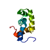
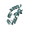
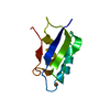
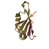
| Entry | Database: PDB / ID: 4y0l | ||||||
|---|---|---|---|---|---|---|---|
| Title | Mycobacterial membrane protein MmpL11D2 | ||||||
 Components Components | Putative membrane protein mmpL11 Biological membrane Biological membrane | ||||||
 Keywords Keywords | MEMBRANE PROTEIN / RND family | ||||||
| Function / homology |  Function and homology information Function and homology information | ||||||
| Biological species |   Mycobacterium tuberculosis (bacteria) Mycobacterium tuberculosis (bacteria) | ||||||
| Method | X-RAY DIFFRACTION / SYNCHROTRON / SAD / Resolution: 2.4 Å | ||||||
 Authors Authors | Torres, R. / Chim, N. / Goulding, C.W. | ||||||
 Citation Citation | Journal: Chem.Biol. / Year: 2015 Title: The Structure and Interactions of Periplasmic Domains of Crucial MmpL Membrane Proteins from Mycobacterium tuberculosis. Authors: Chim, N. / Torres, R. / Liu, Y. / Capri, J. / Batot, G. / Whitelegge, J.P. / Goulding, C.W. | ||||||
| History |
|
- Structure visualization
| Structure viewer | Molecule: MolmilJmol/JSmol |
|---|
- Downloads & links
-Download
| PDBx/mmCIF format | 4y0l.cif.gz | 56.3 KB | Display | PDBx/mmCIF format |
|---|---|---|---|---|
| PDB format | pdb4y0l.ent.gz | 41.4 KB | Display | PDB format |
| PDBx/mmJSON format | 4y0l.json.gz | Tree view | PDBx/mmJSON format | |
| Others |  Other downloads Other downloads |
-Validation report
| Arichive directory | https://data.pdbj.org/pub/pdb/validation_reports/y0/4y0lftp://data.pdbj.org/pub/pdb/validation_reports/y0/4y0l | HTTPS FTP |
|---|
-Related structure data
| Similar structure data |
|---|


-Links
 PDBj
PDBj
- Assembly
| Deposited unit | 
| ||||||||
|---|---|---|---|---|---|---|---|---|---|
| 1 |
| ||||||||
| Unit cell |
| ||||||||
| Components on special symmetry positions |
|
-Components
| #1: Protein | Biological membrane Mass: 9176.288 Da / Num. of mol.: 1 / Fragment: UNP residues 424-511 Source method: isolated from a genetically manipulated source Source: (gene. exp.) Mycobacterium tuberculosis (bacteria) / Strain: ATCC 25618 / H37Rv / Gene: mmpL11, Rv0202c, MTV033.10c / Production host: Escherichia coli (E. coli) / Strain (production host): BL21(DE3) / References: UniProt: P9WJT9 | ||||
|---|---|---|---|---|---|
| #2: Chemical | ChemComp-IOD / Iodide  Mass: 126.904 Da / Num. of mol.: 9 / Source method: obtained synthetically / Formula: I Mass: 126.904 Da / Num. of mol.: 9 / Source method: obtained synthetically / Formula: I#3: Chemical | ChemComp-SO4 / | Sulfate  Mass: 96.063 Da / Num. of mol.: 1 / Source method: obtained synthetically / Formula: SO4 Mass: 96.063 Da / Num. of mol.: 1 / Source method: obtained synthetically / Formula: SO4#4: Water | ChemComp-HOH / | Water Mass: 18.015 Da / Num. of mol.: 16 / Source method: isolated from a natural source / Formula: H2O Mass: 18.015 Da / Num. of mol.: 16 / Source method: isolated from a natural source / Formula: H2O |
-Experimental details
-Experiment
| Experiment | Method: X-RAY DIFFRACTION / Number of used crystals: 1 |
|---|
- Sample preparation
| Crystal | Density Matthews: 2.69 Å3/Da / Density % sol: 54.32 % |
|---|---|
| Crystal grow | Temperature: 293 K / Method: vapor diffusion, hanging drop / pH: 6.7 / Details: 0.1 M MES, pH 6.7, 2 M MgSO4 |
-Data collection
| Diffraction | Mean temperature: 100 K | |||||||||||||||||||||||||||
|---|---|---|---|---|---|---|---|---|---|---|---|---|---|---|---|---|---|---|---|---|---|---|---|---|---|---|---|---|
| Diffraction source | Source: SYNCHROTRON / Site: ALS  / Beamline: 8.2.1 / Wavelength: 1.54 Å / Beamline: 8.2.1 / Wavelength: 1.54 Å | |||||||||||||||||||||||||||
| Detector | Type: ADSC QUANTUM 315r / Detector: CCD / Date: Mar 25, 2014 | |||||||||||||||||||||||||||
| Radiation | Protocol: SINGLE WAVELENGTH / Monochromatic (M) / Laue (L): M / Scattering type: x-ray | |||||||||||||||||||||||||||
| Radiation wavelength | Wavelength: 1.54 Å / Relative weight: 1 | |||||||||||||||||||||||||||
| Reflection | Redundancy: 5.8 % / Number: 23972 / Rmerge(I) obs: 0.109 / D res high: 2.4 Å / D res low: 53.36 Å / Num. obs: 4128 / % possible obs: 99.9 / Rejects: 75 | |||||||||||||||||||||||||||
| Diffraction reflection shell |
| |||||||||||||||||||||||||||
| Reflection | Resolution: 2.4→53.36 Å / Num. obs: 4128 / % possible obs: 99.9 % / Redundancy: 5.8 % / CC1/2: 0.992 / Rmerge(I) obs: 0.109 / Rpim(I) all: 0.048 / Net I/σ(I): 20.5 / Num. measured all: 23972 / Scaling rejects: 75 | |||||||||||||||||||||||||||
| Reflection shell | Diffraction-ID: 1 / Rejects: 0
|
-Phasing
| Phasing | Method: SAD |
|---|---|
| Phasing MAD | D res high: 0 Å / D res low: 0 Å / FOM : 0 / Reflection: 0 |
- Processing
| Software |
| ||||||||||||||||||||||||||||||||||||||||||||||||||||||||||||||||||||||||||||||||||||||||||||||||||||||||||||||||||||||||||||||||||||||||||||||||||||||
|---|---|---|---|---|---|---|---|---|---|---|---|---|---|---|---|---|---|---|---|---|---|---|---|---|---|---|---|---|---|---|---|---|---|---|---|---|---|---|---|---|---|---|---|---|---|---|---|---|---|---|---|---|---|---|---|---|---|---|---|---|---|---|---|---|---|---|---|---|---|---|---|---|---|---|---|---|---|---|---|---|---|---|---|---|---|---|---|---|---|---|---|---|---|---|---|---|---|---|---|---|---|---|---|---|---|---|---|---|---|---|---|---|---|---|---|---|---|---|---|---|---|---|---|---|---|---|---|---|---|---|---|---|---|---|---|---|---|---|---|---|---|---|---|---|---|---|---|---|---|---|---|
| Refinement | Method to determine structure: SAD / Resolution: 2.4→53.359 Å / SU ML: 0.23 / Cross valid method: FREE R-VALUE / σ(F): 1.34 / Phase error: 24.14 / Stereochemistry target values: ML
| ||||||||||||||||||||||||||||||||||||||||||||||||||||||||||||||||||||||||||||||||||||||||||||||||||||||||||||||||||||||||||||||||||||||||||||||||||||||
| Solvent computation | Shrinkage radii: 0.9 Å / VDW probe radii: 1.11 Å / Solvent model: FLAT BULK SOLVENT MODEL | ||||||||||||||||||||||||||||||||||||||||||||||||||||||||||||||||||||||||||||||||||||||||||||||||||||||||||||||||||||||||||||||||||||||||||||||||||||||
| Displacement parameters | Biso max: 104.57 Å2 / Biso mean: 40.1369 Å2 / Biso min: 10.21 Å2 | ||||||||||||||||||||||||||||||||||||||||||||||||||||||||||||||||||||||||||||||||||||||||||||||||||||||||||||||||||||||||||||||||||||||||||||||||||||||
| Refinement step | Cycle: final / Resolution: 2.4→53.359 Å
| ||||||||||||||||||||||||||||||||||||||||||||||||||||||||||||||||||||||||||||||||||||||||||||||||||||||||||||||||||||||||||||||||||||||||||||||||||||||
| Refinement TLS params. | Method: refined / Refine-ID: X-RAY DIFFRACTION
| ||||||||||||||||||||||||||||||||||||||||||||||||||||||||||||||||||||||||||||||||||||||||||||||||||||||||||||||||||||||||||||||||||||||||||||||||||||||
| Refinement TLS group |
|
