 Movie
Movie Controller

[English] 日本語
 Yorodumi
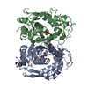
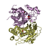
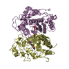
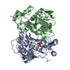
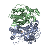
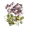
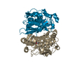
- PDB-4xse: Complex structure of thymidylate synthase from varicella zoster virus -
+ Open data

- Basic information
| Entry | Database: PDB / ID: 4xse | ||||||
|---|---|---|---|---|---|---|---|
| Title | Complex structure of thymidylate synthase from varicella zoster virus | ||||||
 Components Components | Thymidylate synthase | ||||||
 Keywords Keywords | VIRAL PROTEIN / thymidylate synthase / herpesvirus / varicella zoster virus / vzv | ||||||
| Function / homology |  Function and homology informationthymidylate synthase / thymidylate synthase activity / dTMP biosynthetic process / dTTP biosynthetic process / dihydrofolate reductase activity / methylation / cytosol Function and homology informationthymidylate synthase / thymidylate synthase activity / dTMP biosynthetic process / dTTP biosynthetic process / dihydrofolate reductase activity / methylation / cytosolSimilarity search - Function | ||||||
| Biological species | Varicella-zoster virus | ||||||
| Method | X-RAY DIFFRACTION / SYNCHROTRON / MOLECULAR REPLACEMENT / Resolution: 3.1 Å | ||||||
 Authors Authors | Hew, K. | ||||||
 Citation Citation | Journal: Plos One / Year: 2015 Title: Structure of the Varicella Zoster Virus Thymidylate Synthase Establishes Functional and Structural Similarities as the Human Enzyme and Potentiates Itself as a Target of Brivudine. Authors: Hew, K. / Dahlroth, S.L. / Veerappan, S. / Pan, L.X. / Cornvik, T. / Nordlund, P. | ||||||
| History |
|
- Structure visualization
| Structure viewer | Molecule: MolmilJmol/JSmol |
|---|
- Downloads & links
-Download
| PDBx/mmCIF format | 4xse.cif.gz | 229.4 KB | Display | PDBx/mmCIF format |
|---|---|---|---|---|
| PDB format | pdb4xse.ent.gz | 186.8 KB | Display | PDB format |
| PDBx/mmJSON format | 4xse.json.gz | Tree view | PDBx/mmJSON format | |
| Others |  Other downloads Other downloads |
-Validation report
| Arichive directory | https://data.pdbj.org/pub/pdb/validation_reports/xs/4xseftp://data.pdbj.org/pub/pdb/validation_reports/xs/4xse | HTTPS FTP |
|---|
-Related structure data
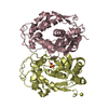
| Related structure data | 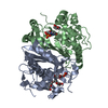 4xscC 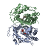 4xsdC  1hzwS S: Starting model for refinement C: citing same article ( |
|---|---|
| Similar structure data |


-Links
 PDBj
PDBj- Assembly
| Deposited unit | 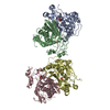
| ||||||||
|---|---|---|---|---|---|---|---|---|---|
| 1 | 
| ||||||||
| 2 | 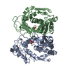
| ||||||||
| Unit cell |
|
-Components
| #1: Protein | / TSase Mass: 35768.848 Da / Num. of mol.: 4 Source method: isolated from a genetically manipulated source Source: (gene. exp.)  Varicella-zoster virus (strain Oka vaccine) Varicella-zoster virus (strain Oka vaccine)Gene: ORF13 / Production host:  Escherichia coli (E. coli) / References: UniProt: Q4JQW2, thymidylate synthase Escherichia coli (E. coli) / References: UniProt: Q4JQW2, thymidylate synthase#2: Chemical | ChemComp-PO4 / Phosphate  Mass: 94.971 Da / Num. of mol.: 4 / Source method: obtained synthetically / Formula: PO4 Mass: 94.971 Da / Num. of mol.: 4 / Source method: obtained synthetically / Formula: PO4 |
|---|
-Experimental details
-Experiment
| Experiment | Method: X-RAY DIFFRACTION |
|---|
- Sample preparation
| Crystal | Density Matthews: 4.23 Å3/Da / Density % sol: 70.94 % |
|---|---|
| Crystal grow | Temperature: 293 K / Method: vapor diffusion, sitting drop / Details: 12% ethylene glycol, 0.1 M HEPES pH 7.5 |
-Data collection
| Diffraction | Mean temperature: 100 K |
|---|---|
| Diffraction source | Source: SYNCHROTRON / Site: Australian Synchrotron  / Beamline: MX1 / Wavelength: 0.9537 Å / Beamline: MX1 / Wavelength: 0.9537 Å |
| Detector | Type: ADSC QUANTUM 210r / Detector: CCD / Date: Mar 27, 2014 |
| Radiation | Protocol: SINGLE WAVELENGTH / Monochromatic (M) / Laue (L): M / Scattering type: x-ray |
| Radiation wavelength | Wavelength: 0.9537 Å / Relative weight: 1 |
| Reflection | Resolution: 3.1→29.8 Å / Num. obs: 42243 / % possible obs: 99.2 % / Redundancy: 3.7 % / Rmerge(I) obs: 0.127 / Net I/σ(I): 8.5 |
| Reflection shell | Resolution: 3.1→3.2 Å / Redundancy: 3.4 % / Rmerge(I) obs: 0.475 / % possible all: 97.5 |
- Processing
| Software |
| |||||||||||||||||||||||||||||||||||||||||||||||||||||||||||||||||||||||||||||||||||||||||||||||||||||||||
|---|---|---|---|---|---|---|---|---|---|---|---|---|---|---|---|---|---|---|---|---|---|---|---|---|---|---|---|---|---|---|---|---|---|---|---|---|---|---|---|---|---|---|---|---|---|---|---|---|---|---|---|---|---|---|---|---|---|---|---|---|---|---|---|---|---|---|---|---|---|---|---|---|---|---|---|---|---|---|---|---|---|---|---|---|---|---|---|---|---|---|---|---|---|---|---|---|---|---|---|---|---|---|---|---|---|---|
| Refinement | Method to determine structure: MOLECULAR REPLACEMENT Starting model: 1HZW Resolution: 3.1→29.727 Å / Cross valid method: FREE R-VALUE / σ(F): 1.98 / Phase error: 35.83 / Stereochemistry target values: TWIN_LSQ_F
| |||||||||||||||||||||||||||||||||||||||||||||||||||||||||||||||||||||||||||||||||||||||||||||||||||||||||
| Solvent computation | Shrinkage radii: 0.9 Å / VDW probe radii: 1.11 Å / Solvent model: FLAT BULK SOLVENT MODEL | |||||||||||||||||||||||||||||||||||||||||||||||||||||||||||||||||||||||||||||||||||||||||||||||||||||||||
| Refinement step | Cycle: LAST / Resolution: 3.1→29.727 Å
| |||||||||||||||||||||||||||||||||||||||||||||||||||||||||||||||||||||||||||||||||||||||||||||||||||||||||
| Refine LS restraints |
| |||||||||||||||||||||||||||||||||||||||||||||||||||||||||||||||||||||||||||||||||||||||||||||||||||||||||
| LS refinement shell |
|
