 Movie
Movie Controller

[English] 日本語
 Yorodumi
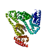
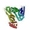
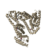
- PDB-4xs0: Human methemoglobin in complex with the second and third NEAT dom... -
+ Open data

- Basic information
| Entry | Database: PDB / ID: 4xs0 | ||||||
|---|---|---|---|---|---|---|---|
| Title | Human methemoglobin in complex with the second and third NEAT domains of IsdH(F365Y/A369F/Y642A) from Staphylococcus aureus | ||||||
 Components Components |
| ||||||
 Keywords Keywords | METAL TRANSPORT / NEAT / HEME/HEMOGLOBIN BINDING /  HEMOGLOBIN / OXYGEN TRANSPORT-PROTEIN BINDING COMPLEX / oxygen transport HEMOGLOBIN / OXYGEN TRANSPORT-PROTEIN BINDING COMPLEX / oxygen transport | ||||||
| Function / homology |  Function and homology information Function and homology informationcellular oxidant detoxification / nitric oxide transport / hemoglobin alpha binding / haptoglobin-hemoglobin complex / hemoglobin binding / organic acid binding / renal absorption / hemoglobin complex / oxygen transport / Scavenging of heme from plasma ...cellular oxidant detoxification / nitric oxide transport / hemoglobin alpha binding / haptoglobin-hemoglobin complex / hemoglobin binding / organic acid binding / renal absorption / hemoglobin complex / oxygen transport / Scavenging of heme from plasma / endocytic vesicle lumen / blood vessel diameter maintenance / hydrogen peroxide catabolic process / oxygen carrier activity / Late endosomal microautophagy / Heme signaling / carbon dioxide transport / Erythrocytes take up oxygen and release carbon dioxide / response to hydrogen peroxide / Erythrocytes take up carbon dioxide and release oxygen / Cytoprotection by HMOX1 / platelet aggregation / oxygen binding / regulation of blood pressure / Chaperone Mediated Autophagy / positive regulation of nitric oxide biosynthetic process / tertiary granule lumen / Factors involved in megakaryocyte development and platelet production / ficolin-1-rich granule lumen / blood microparticle / iron ion binding / heme binding / Neutrophil degranulation / extracellular space / extracellular exosome / extracellular region / membrane / metal ion binding / cytosolSimilarity search - Function | ||||||
| Biological species |   Staphylococcus aureus (bacteria) Staphylococcus aureus (bacteria) Homo sapiens (human) Homo sapiens (human) | ||||||
| Method | X-RAY DIFFRACTION / SYNCHROTRON / MOLECULAR REPLACEMENT / molecular replacement / Resolution: 2.55 Å | ||||||
 Authors Authors | Dickson, C.F. / Jacques, D.A. / Guss, J.M. / Gell, D.A. | ||||||
 Citation Citation | Journal: Acta Crystallogr.,Sect.D / Year: 2015 Title: The structure of haemoglobin bound to the haemoglobin receptor IsdH from Staphylococcus aureus shows disruption of the native alpha-globin haem pocket. Authors: Dickson, C.F. / Jacques, D.A. / Clubb, R.T. / Guss, J.M. / Gell, D.A. | ||||||
| History |
|
- Structure visualization
| Structure viewer | Molecule: MolmilJmol/JSmol |
|---|
- Downloads & links
-Download
| PDBx/mmCIF format | 4xs0.cif.gz | 256.1 KB | Display | PDBx/mmCIF format |
|---|---|---|---|---|
| PDB format | pdb4xs0.ent.gz | 206.6 KB | Display | PDB format |
| PDBx/mmJSON format | 4xs0.json.gz | Tree view | PDBx/mmJSON format | |
| Others |  Other downloads Other downloads |
-Validation report
| Arichive directory | https://data.pdbj.org/pub/pdb/validation_reports/xs/4xs0ftp://data.pdbj.org/pub/pdb/validation_reports/xs/4xs0 | HTTPS FTP |
|---|
-Related structure data
| Related structure data |  4ij2S S: Starting model for refinement |
|---|---|
| Similar structure data |


-Links
 PDBj
PDBj


- Assembly
| Deposited unit | 
| ||||||||
|---|---|---|---|---|---|---|---|---|---|
| 1 |
| ||||||||
| Unit cell |
|
-Components
-Hemoglobin subunit ... , 2 types, 2 molecules AB
| #1: Protein | / Alpha-globin / Hemoglobin alpha chain Mass: 15150.353 Da / Num. of mol.: 1 / Source method: isolated from a natural source / Source: (natural) Homo sapiens (human) / References: UniProt: P69905 |
|---|---|
| #2: Protein | / Beta-globin / Hemoglobin beta chain Mass: 15890.198 Da / Num. of mol.: 1 / Source method: isolated from a natural source / Source: (natural) Homo sapiens (human) / References: UniProt: P68871 |
-Protein , 1 types, 1 molecules C
| #3: Protein | Mass: 38929.402 Da / Num. of mol.: 1 / Fragment: UNP residues 326-660 / Mutation: F365Y/A369F/Y642A Source method: isolated from a genetically manipulated source Source: (gene. exp.) Staphylococcus aureus (bacteria) / Strain: USA300 / Gene: isdH, harA, sasI, SAUSA300_1677 / Production host: Escherichia coli BL21(DE3) (bacteria) / Variant (production host): Rosetta 2 / References: UniProt: Q2FG07 |
|---|
-Non-polymers , 4 types, 10 molecules 


| #4: Chemical | Heme B Mass: 616.487 Da / Num. of mol.: 2 / Source method: obtained synthetically / Formula: C34H32FeN4O4 Mass: 616.487 Da / Num. of mol.: 2 / Source method: obtained synthetically / Formula: C34H32FeN4O4#5: Chemical | Chloride Mass: 35.453 Da / Num. of mol.: 3 / Source method: obtained synthetically / Formula: Cl Mass: 35.453 Da / Num. of mol.: 3 / Source method: obtained synthetically / Formula: Cl#6: Chemical | ChemComp-SO4 / | Sulfate Mass: 96.063 Da / Num. of mol.: 1 / Source method: obtained synthetically / Formula: SO4 Mass: 96.063 Da / Num. of mol.: 1 / Source method: obtained synthetically / Formula: SO4#7: Water | ChemComp-HOH / | WaterMass: 18.015 Da / Num. of mol.: 4 / Source method: isolated from a natural source / Formula: H2O |
|---|
-Experimental details
-Experiment
| Experiment | Method: X-RAY DIFFRACTION / Number of used crystals: 1 |
|---|
- Sample preparation
| Crystal | Density Matthews: 3.2 Å3/Da / Density % sol: 61.59 % |
|---|---|
| Crystal grow | Temperature: 294 K / Method: vapor diffusion, hanging drop Details: 0.2 M K/Na tartrate, 0.1 M tri-sodium citrate pH 5.6, 2 M ammonium sulfate. Cryoprotected in 20% glycerol. PH range: 5.4 - 5.8 |
-Data collection
| Diffraction | Mean temperature: 100 K | |||||||||||||||||||||||||||
|---|---|---|---|---|---|---|---|---|---|---|---|---|---|---|---|---|---|---|---|---|---|---|---|---|---|---|---|---|
| Diffraction source | Source: SYNCHROTRON / Site: Australian Synchrotron  / Beamline: MX1 / Wavelength: 0.9658 Å / Beamline: MX1 / Wavelength: 0.9658 Å | |||||||||||||||||||||||||||
| Detector | Type: ADSC QUANTUM 210r / Detector: CCD / Date: Nov 15, 2013 | |||||||||||||||||||||||||||
| Radiation | Monochromator: Si(111) / Protocol: SINGLE WAVELENGTH / Monochromatic (M) / Laue (L): M / Scattering type: x-ray | |||||||||||||||||||||||||||
| Radiation wavelength | Wavelength: 0.9658 Å / Relative weight: 1 | |||||||||||||||||||||||||||
| Reflection | Resolution: 2.55→33.39 Å / Num. obs: 31237 / % possible obs: 99.9 % / Redundancy: 9.6 % / CC1/2: 0.996 / Rmerge(I) obs: 0.138 / Rpim(I) all: 0.042 / Net I/σ(I): 9.3 / Num. measured all: 298616 / Scaling rejects: 181 | |||||||||||||||||||||||||||
| Reflection shell | Diffraction-ID: 1 / Rejects: 0
|
-Phasing
| Phasing | Method: molecular replacement | |||||||||
|---|---|---|---|---|---|---|---|---|---|---|
| Phasing MR | Model details: Phaser MODE: MR_AUTO
|
- Processing
| Software |
| |||||||||||||||||||||||||||||||||||||||||||||||||||||||||||||||||||||||||||||||||||||||||||||||||||||||||||||||||||||||||||||
|---|---|---|---|---|---|---|---|---|---|---|---|---|---|---|---|---|---|---|---|---|---|---|---|---|---|---|---|---|---|---|---|---|---|---|---|---|---|---|---|---|---|---|---|---|---|---|---|---|---|---|---|---|---|---|---|---|---|---|---|---|---|---|---|---|---|---|---|---|---|---|---|---|---|---|---|---|---|---|---|---|---|---|---|---|---|---|---|---|---|---|---|---|---|---|---|---|---|---|---|---|---|---|---|---|---|---|---|---|---|---|---|---|---|---|---|---|---|---|---|---|---|---|---|---|---|---|
| Refinement | Method to determine structure: MOLECULAR REPLACEMENT Starting model: 4IJ2 Resolution: 2.55→33.39 Å / Cor.coef. Fo:Fc: 0.918 / Cor.coef. Fo:Fc free: 0.888 / WRfactor Rfree: 0.2675 / WRfactor Rwork: 0.2428 / FOM work R set: 0.7377 / SU B: 33.489 / SU ML: 0.303 / SU R Cruickshank DPI: 0.4685 / SU Rfree: 0.3027 / Cross valid method: THROUGHOUT / σ(F): 0 / ESU R: 0.469 / ESU R Free: 0.303 / Stereochemistry target values: MAXIMUM LIKELIHOOD Details: HYDROGENS HAVE BEEN ADDED IN THE RIDING POSITIONS U VALUES : WITH TLS ADDED
| |||||||||||||||||||||||||||||||||||||||||||||||||||||||||||||||||||||||||||||||||||||||||||||||||||||||||||||||||||||||||||||
| Solvent computation | Ion probe radii: 0.7 Å / Shrinkage radii: 0.7 Å / VDW probe radii: 1 Å / Solvent model: MASK | |||||||||||||||||||||||||||||||||||||||||||||||||||||||||||||||||||||||||||||||||||||||||||||||||||||||||||||||||||||||||||||
| Displacement parameters | Biso max: 138.74 Å2 / Biso mean: 67.181 Å2 / Biso min: 28.06 Å2
| |||||||||||||||||||||||||||||||||||||||||||||||||||||||||||||||||||||||||||||||||||||||||||||||||||||||||||||||||||||||||||||
| Refinement step | Cycle: final / Resolution: 2.55→33.39 Å
| |||||||||||||||||||||||||||||||||||||||||||||||||||||||||||||||||||||||||||||||||||||||||||||||||||||||||||||||||||||||||||||
| Refine LS restraints |
| |||||||||||||||||||||||||||||||||||||||||||||||||||||||||||||||||||||||||||||||||||||||||||||||||||||||||||||||||||||||||||||
| LS refinement shell | Resolution: 2.55→2.616 Å / Total num. of bins used: 20
| |||||||||||||||||||||||||||||||||||||||||||||||||||||||||||||||||||||||||||||||||||||||||||||||||||||||||||||||||||||||||||||
| Refinement TLS params. | Method: refined / Refine-ID: X-RAY DIFFRACTION
| |||||||||||||||||||||||||||||||||||||||||||||||||||||||||||||||||||||||||||||||||||||||||||||||||||||||||||||||||||||||||||||
| Refinement TLS group |
|
