 Movie
Movie Controller


+ Open data

- Basic information
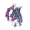
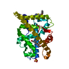
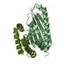
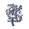
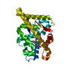
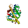
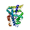
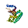
| Entry | Database: PDB / ID: 4xiz | ||||||
|---|---|---|---|---|---|---|---|
| Title | Structure of a phospholipid trafficking complex with substrate | ||||||
 Components Components |
| ||||||
 Keywords Keywords | LIPID TRANSPORT/OXIDOREDUCTASE /  phospholipid / LIPID TRANSPORT-OXIDOREDUCTASE complex phospholipid / LIPID TRANSPORT-OXIDOREDUCTASE complex | ||||||
| Function / homology |  Function and homology information Function and homology informationTP53 Regulates Transcription of Genes Involved in Cytochrome C Release / phosphatidic acid transfer activity / cardiolipin metabolic process / positive regulation of phosphatidylcholine biosynthetic process / mitochondrial respiratory chain complex assembly / phospholipid transport / phospholipid translocation / mitochondrion organization / mitochondrial intermembrane space / mitochondrial inner membrane ...TP53 Regulates Transcription of Genes Involved in Cytochrome C Release / phosphatidic acid transfer activity / cardiolipin metabolic process / positive regulation of phosphatidylcholine biosynthetic process / mitochondrial respiratory chain complex assembly / phospholipid transport / phospholipid translocation / mitochondrion organization / mitochondrial intermembrane space / mitochondrial inner membrane / lipid binding / mitochondrion / nucleus / cytosol / cytoplasmSimilarity search - Function | ||||||
| Biological species |  Saccharomyces cerevisiae (brewer's yeast) Saccharomyces cerevisiae (brewer's yeast) | ||||||
| Method | X-RAY DIFFRACTION / SYNCHROTRON / SAD / Resolution: 2 Å | ||||||
 Authors Authors | Yu, F. / He, F. / Wang, C. / Zhang, P. | ||||||
 Citation Citation | Journal: Embo Rep. / Year: 2015 Title: Structural basis of intramitochondrial phosphatidic acid transport mediated by Ups1-Mdm35 complex Authors: Yu, F. / He, F. / Yao, H. / Wang, C. / Wang, J. / Li, J. / Qi, X. / Xue, H. / Ding, J. / Zhang, P. | ||||||
| History |
|
- Structure visualization
| Structure viewer | Molecule: MolmilJmol/JSmol |
|---|
- Downloads & links
-Download
| PDBx/mmCIF format | 4xiz.cif.gz | 115.1 KB | Display | PDBx/mmCIF format |
|---|---|---|---|---|
| PDB format | pdb4xiz.ent.gz | 93.8 KB | Display | PDB format |
| PDBx/mmJSON format | 4xiz.json.gz | Tree view | PDBx/mmJSON format | |
| Others |  Other downloads Other downloads |
-Validation report
| Arichive directory | https://data.pdbj.org/pub/pdb/validation_reports/xi/4xizftp://data.pdbj.org/pub/pdb/validation_reports/xi/4xiz | HTTPS FTP |
|---|
-Related structure data


-Links
 PDBj
PDBj

- Assembly
| Deposited unit | 
| ||||||||
|---|---|---|---|---|---|---|---|---|---|
| 1 | 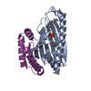
| ||||||||
| 2 | 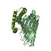
| ||||||||
| Unit cell |
|
-Components
| #1: Protein | Mass: 19493.252 Da / Num. of mol.: 2 Source method: isolated from a genetically manipulated source Source: (gene. exp.) Saccharomyces cerevisiae (strain ATCC 204508 / S288c) (yeast)Strain: ATCC 204508 / S288c / Gene: UPS1, YLR193C / Production host:  Escherichia coli (E. coli) / References: UniProt: Q05776 Escherichia coli (E. coli) / References: UniProt: Q05776#2: Protein | Mass: 7975.900 Da / Num. of mol.: 2 / Fragment: UNP residues 6-75 Source method: isolated from a genetically manipulated source Source: (gene. exp.) Saccharomyces cerevisiae (strain ATCC 204508 / S288c) (yeast)Strain: ATCC 204508 / S288c / Gene: MDM35, YKL053C-A / Production host: Escherichia coli (E. coli) / References: UniProt: O60200#3: Chemical |   Mass: 648.891 Da / Num. of mol.: 2 / Source method: obtained synthetically / Formula: C35H69O8P / Comment: phospholipid*YM Mass: 648.891 Da / Num. of mol.: 2 / Source method: obtained synthetically / Formula: C35H69O8P / Comment: phospholipid*YM#4: Water | ChemComp-HOH / | Water Mass: 18.015 Da / Num. of mol.: 380 / Source method: isolated from a natural source / Formula: H2O Mass: 18.015 Da / Num. of mol.: 380 / Source method: isolated from a natural source / Formula: H2O |
|---|
-Experimental details
-Experiment
| Experiment | Method: X-RAY DIFFRACTION |
|---|
- Sample preparation
| Crystal | Density Matthews: 2.54 Å3/Da / Density % sol: 51.63 % |
|---|---|
| Crystal grow | Temperature: 277.15 K / Method: vapor diffusion, sitting drop Details: 2%(v/v)Tacsimate pH 6.0, 0.1M Bis-Tris pH 6.5, 20%(w/v) PEG3350 |
-Data collection
| Diffraction | Mean temperature: 100 K | ||||||||||||||||||||||||||||||||||||||||||||||||||||||||||||||||||
|---|---|---|---|---|---|---|---|---|---|---|---|---|---|---|---|---|---|---|---|---|---|---|---|---|---|---|---|---|---|---|---|---|---|---|---|---|---|---|---|---|---|---|---|---|---|---|---|---|---|---|---|---|---|---|---|---|---|---|---|---|---|---|---|---|---|---|---|
| Diffraction source | Source: SYNCHROTRON / Site: SSRF  / Beamline: BL17U / Wavelength: 0.9793 Å / Beamline: BL17U / Wavelength: 0.9793 Å | ||||||||||||||||||||||||||||||||||||||||||||||||||||||||||||||||||
| Detector | Type: MARMOSAIC 225 mm CCD / Detector: CCD / Date: Jun 29, 2013 | ||||||||||||||||||||||||||||||||||||||||||||||||||||||||||||||||||
| Radiation | Protocol: SINGLE WAVELENGTH / Monochromatic (M) / Laue (L): M / Scattering type: x-ray | ||||||||||||||||||||||||||||||||||||||||||||||||||||||||||||||||||
| Radiation wavelength | Wavelength: 0.9793 Å / Relative weight: 1 | ||||||||||||||||||||||||||||||||||||||||||||||||||||||||||||||||||
| Reflection | Resolution: 2→50 Å / Num. obs: 36740 / % possible obs: 99.4 % / Redundancy: 4.1 % / Rmerge(I) obs: 0.116 / Χ2: 1.394 / Net I/av σ(I): 15.354 / Net I/σ(I): 10.7 / Num. measured all: 150394 | ||||||||||||||||||||||||||||||||||||||||||||||||||||||||||||||||||
| Reflection shell | Diffraction-ID: 1 / Rejects: 0
|
- Processing
| Software |
| ||||||||||||||||
|---|---|---|---|---|---|---|---|---|---|---|---|---|---|---|---|---|---|
| Refinement | Method to determine structure: SAD / Resolution: 2→34.13 Å / Cross valid method: FREE R-VALUE
| ||||||||||||||||
| Displacement parameters | Biso max: 137.57 Å2 / Biso mean: 43.322 Å2 / Biso min: 16.54 Å2 | ||||||||||||||||
| Refinement step | Cycle: LAST / Resolution: 2→34.13 Å
|
