 Movie
Movie Controller

[English] 日本語
 Yorodumi
- PDB-4xep: Crystal Structure of F222 form of E112A/H234A Mutant of Stationar... -
+ Open data

- Basic information
| Entry | Database: PDB / ID: 4xep | ||||||
|---|---|---|---|---|---|---|---|
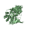
| Title | Crystal Structure of F222 form of E112A/H234A Mutant of Stationary Phase Survival Protein (SurE) from Salmonella typhimurium | ||||||
 Components Components | 5'/3'-nucleotidase SurE | ||||||
 Keywords Keywords |  HYDROLASE / Stationary phase survival protein / Domain swapping / Rossmann fold like / Phosphatase HYDROLASE / Stationary phase survival protein / Domain swapping / Rossmann fold like / Phosphatase | ||||||
| Function / homology |  Function and homology information3'-nucleotidase / 3'-nucleotidase activity / exopolyphosphatase / exopolyphosphatase activity / XMP 5'-nucleosidase activity / 5'-nucleotidase / 5'-nucleotidase activity / nucleotide binding / metal ion binding / cytoplasm Function and homology information3'-nucleotidase / 3'-nucleotidase activity / exopolyphosphatase / exopolyphosphatase activity / XMP 5'-nucleosidase activity / 5'-nucleotidase / 5'-nucleotidase activity / nucleotide binding / metal ion binding / cytoplasmSimilarity search - Function | ||||||
| Biological species |  Salmonella typhimurium LT2 (bacteria) Salmonella typhimurium LT2 (bacteria) | ||||||
| Method | X-RAY DIFFRACTION / SYNCHROTRON / MOLECULAR REPLACEMENT / Resolution: 1.5 Å | ||||||
 Authors Authors | Mathiharan, Y.K. / Murthy, M.R.N. | ||||||
 Citation Citation | Journal: Acta Crystallogr.,Sect.D / Year: 2015 Title: Insights into stabilizing interactions in the distorted domain-swapped dimer of Salmonella typhimurium survival protein. Authors: Mathiharan, Y.K. / Savithri, H.S. / Murthy, M.R. | ||||||
| History |
|
- Structure visualization
| Structure viewer | Molecule: MolmilJmol/JSmol |
|---|
- Downloads & links
-Download
| PDBx/mmCIF format | 4xep.cif.gz | 133 KB | Display | PDBx/mmCIF format |
|---|---|---|---|---|
| PDB format | pdb4xep.ent.gz | 102 KB | Display | PDB format |
| PDBx/mmJSON format | 4xep.json.gz | Tree view | PDBx/mmJSON format | |
| Others |  Other downloads Other downloads |
-Validation report
| Arichive directory | https://data.pdbj.org/pub/pdb/validation_reports/xe/4xepftp://data.pdbj.org/pub/pdb/validation_reports/xe/4xep | HTTPS FTP |
|---|
-Related structure data
| Related structure data |  4rytC  4ryuC  4xerC 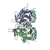 4xgbC  4xgpC  4xh8C  4xj7C  2v4nS C: citing same article ( S: Starting model for refinement |
|---|---|
| Similar structure data |


-Links
 PDBj
PDBj- Assembly
| Deposited unit | 
| |||||||||||||||||||||||||||
|---|---|---|---|---|---|---|---|---|---|---|---|---|---|---|---|---|---|---|---|---|---|---|---|---|---|---|---|---|
| 1 | 
| |||||||||||||||||||||||||||
| Unit cell |
| |||||||||||||||||||||||||||
| Components on special symmetry positions |
|
-Components
| #1: Protein | Mass: 28490.008 Da / Num. of mol.: 1 / Mutation: E112A, H234A Source method: isolated from a genetically manipulated source Source: (gene. exp.) Salmonella typhimurium LT2 (bacteria) / Strain: LT2 / Gene: surE / Plasmid: pRSETC / Production host: Escherichia coli (E. coli) / Strain (production host): BL21-pLysSReferences: UniProt: P66881, 5'-nucleotidase, 3'-nucleotidase, exopolyphosphatase | ||
|---|---|---|---|
| #2: Chemical | ChemComp-MG /   Mass: 24.305 Da / Num. of mol.: 1 / Source method: obtained synthetically / Formula: Mg Mass: 24.305 Da / Num. of mol.: 1 / Source method: obtained synthetically / Formula: Mg | ||
| #3: Chemical | ChemComp-PO4 / Phosphate  Mass: 94.971 Da / Num. of mol.: 1 / Source method: obtained synthetically / Formula: PO4 Mass: 94.971 Da / Num. of mol.: 1 / Source method: obtained synthetically / Formula: PO4 | ||
| #4: Chemical | ChemComp-EDO / Ethylene glycol  Mass: 62.068 Da / Num. of mol.: 4 / Source method: obtained synthetically / Formula: C2H6O2 Mass: 62.068 Da / Num. of mol.: 4 / Source method: obtained synthetically / Formula: C2H6O2#5: Water | ChemComp-HOH / | Water Mass: 18.015 Da / Num. of mol.: 353 / Source method: isolated from a natural source / Formula: H2O Mass: 18.015 Da / Num. of mol.: 353 / Source method: isolated from a natural source / Formula: H2O |
-Experimental details
-Experiment
| Experiment | Method: X-RAY DIFFRACTION |
|---|
- Sample preparation
| Crystal | Density Matthews: 2.81 Å3/Da / Density % sol: 56.3 % |
|---|---|
| Crystal grow | Temperature: 293 K / Method: microbatch / pH: 7.5 Details: 0.1 M HEPES pH 7.5, 0.02 M MgCl2 hexahydrate, 22% (w/v) Polyacrylic acid 5100 sodium salt |
-Data collection
| Diffraction | Mean temperature: 100 K |
|---|---|
| Diffraction source | Source: SYNCHROTRON / Site: ESRF  / Beamline: BM14 / Wavelength: 0.97625 Å / Beamline: BM14 / Wavelength: 0.97625 Å |
| Detector | Type: MARMOSAIC 225 mm CCD / Detector: CCD / Date: Apr 22, 2013 / Details: bent collimating mirror and toroid |
| Radiation | Monochromator: Si(111) monochromator / Protocol: SINGLE WAVELENGTH / Monochromatic (M) / Laue (L): M / Scattering type: x-ray |
| Radiation wavelength | Wavelength: 0.97625 Å / Relative weight: 1 |
| Reflection | Resolution: 1.5→50 Å / Num. obs: 49090 / % possible obs: 95.5 % / Redundancy: 9.7 % / Biso Wilson estimate: 24.1 Å2 / Rsym value: 0.074 / Net I/σ(I): 68.1 |
| Reflection shell | Resolution: 1.5→1.53 Å / Redundancy: 4.1 % / Rmerge(I) obs: 0.479 / Mean I/σ(I) obs: 2.4 / % possible all: 75.4 |
- Processing
| Software |
| ||||||||||||||||||||||||||||||||||||||||||||||||||||||||||||||||||||||||||||||||||||||||||||||||||||||||||||||||||||||||||||||||||||||||||||||||||||||||||||||||||||||||||||||||||||||
|---|---|---|---|---|---|---|---|---|---|---|---|---|---|---|---|---|---|---|---|---|---|---|---|---|---|---|---|---|---|---|---|---|---|---|---|---|---|---|---|---|---|---|---|---|---|---|---|---|---|---|---|---|---|---|---|---|---|---|---|---|---|---|---|---|---|---|---|---|---|---|---|---|---|---|---|---|---|---|---|---|---|---|---|---|---|---|---|---|---|---|---|---|---|---|---|---|---|---|---|---|---|---|---|---|---|---|---|---|---|---|---|---|---|---|---|---|---|---|---|---|---|---|---|---|---|---|---|---|---|---|---|---|---|---|---|---|---|---|---|---|---|---|---|---|---|---|---|---|---|---|---|---|---|---|---|---|---|---|---|---|---|---|---|---|---|---|---|---|---|---|---|---|---|---|---|---|---|---|---|---|---|---|---|
| Refinement | Method to determine structure: MOLECULAR REPLACEMENT Starting model: 2V4N Resolution: 1.5→50 Å / Cor.coef. Fo:Fc: 0.971 / Cor.coef. Fo:Fc free: 0.956 / SU B: 3.08 / SU ML: 0.058 / Cross valid method: THROUGHOUT / ESU R: 0.076 / ESU R Free: 0.082 / Stereochemistry target values: MAXIMUM LIKELIHOOD / Details: HYDROGENS HAVE BEEN USED IF PRESENT IN THE INPUT
| ||||||||||||||||||||||||||||||||||||||||||||||||||||||||||||||||||||||||||||||||||||||||||||||||||||||||||||||||||||||||||||||||||||||||||||||||||||||||||||||||||||||||||||||||||||||
| Solvent computation | Ion probe radii: 0.8 Å / Shrinkage radii: 0.8 Å / VDW probe radii: 1.2 Å / Solvent model: MASK | ||||||||||||||||||||||||||||||||||||||||||||||||||||||||||||||||||||||||||||||||||||||||||||||||||||||||||||||||||||||||||||||||||||||||||||||||||||||||||||||||||||||||||||||||||||||
| Displacement parameters | Biso mean: 32.593 Å2
| ||||||||||||||||||||||||||||||||||||||||||||||||||||||||||||||||||||||||||||||||||||||||||||||||||||||||||||||||||||||||||||||||||||||||||||||||||||||||||||||||||||||||||||||||||||||
| Refinement step | Cycle: 1 / Resolution: 1.5→50 Å
| ||||||||||||||||||||||||||||||||||||||||||||||||||||||||||||||||||||||||||||||||||||||||||||||||||||||||||||||||||||||||||||||||||||||||||||||||||||||||||||||||||||||||||||||||||||||
| Refine LS restraints |
|