 Movie
Movie Controller

[English] 日本語
 Yorodumi
- PDB-4xcy: Crystal structure of human 4E10 Fab in complex with phosphatidylg... -
+ Open data

- Basic information
| Entry | Database: PDB / ID: 4xcy | ||||||
|---|---|---|---|---|---|---|---|
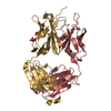
| Title | Crystal structure of human 4E10 Fab in complex with phosphatidylglycerol (06:0 PG) | ||||||
 Components Components |
| ||||||
 Keywords Keywords |  IMMUNE SYSTEM / 4E10 Fab anti HIV-1 gp41 MPER / membrane lipids / phosphatidylglycerol IMMUNE SYSTEM / 4E10 Fab anti HIV-1 gp41 MPER / membrane lipids / phosphatidylglycerol | ||||||
| Function / homology | Immunoglobulins / Immunoglobulin-like / Sandwich / Mainly Beta / Chem-44G / Unknown ligand Function and homology information Function and homology information | ||||||
| Biological species |  Homo sapiens (human) Homo sapiens (human) | ||||||
| Method | X-RAY DIFFRACTION / SYNCHROTRON / MOLECULAR REPLACEMENT / Resolution: 3.96 Å | ||||||
 Authors Authors | Irimia, A. / Stanfield, R.L. / Wilson, I.A. | ||||||
| Funding support |  United States, 1items United States, 1items
| ||||||
 Citation Citation | Journal: Immunity / Year: 2016 Title: Crystallographic Identification of Lipid as an Integral Component of the Epitope of HIV Broadly Neutralizing Antibody 4E10. Authors: Irimia, A. / Sarkar, A. / Stanfield, R.L. / Wilson, I.A. | ||||||
| History |
|
- Structure visualization
| Structure viewer | Molecule: MolmilJmol/JSmol |
|---|
- Downloads & links
-Download
| PDBx/mmCIF format | 4xcy.cif.gz | 436.1 KB | Display | PDBx/mmCIF format |
|---|---|---|---|---|
| PDB format | pdb4xcy.ent.gz | 359.7 KB | Display | PDB format |
| PDBx/mmJSON format | 4xcy.json.gz | Tree view | PDBx/mmJSON format | |
| Others |  Other downloads Other downloads |
-Validation report
| Arichive directory | https://data.pdbj.org/pub/pdb/validation_reports/xc/4xcyftp://data.pdbj.org/pub/pdb/validation_reports/xc/4xcy | HTTPS FTP |
|---|
-Related structure data
| Related structure data | 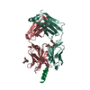 4xawC 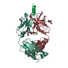 4xbeC  4xbgC  4xbpC 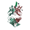 4xc1C 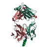 4xc3C 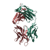 4xccC 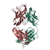 4xceC 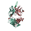 4xcfC  4xcnC 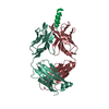 2fx7S C: citing same article ( S: Starting model for refinement |
|---|---|
| Similar structure data |

-Links
 PDBj
PDBj


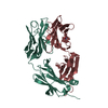
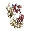
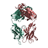
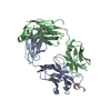
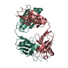
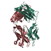
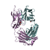
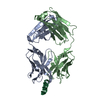
- Assembly
| Deposited unit | 
| ||||||||
|---|---|---|---|---|---|---|---|---|---|
| 1 | 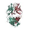
| ||||||||
| 2 | 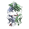
| ||||||||
| 3 | 
| ||||||||
| 4 | 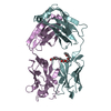
| ||||||||
| 5 | 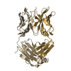
| ||||||||
| 6 | 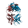
| ||||||||
| Unit cell |
|
-Components
| #1: Antibody | Mass: 23395.850 Da / Num. of mol.: 6 Source method: isolated from a genetically manipulated source Source: (gene. exp.) Homo sapiens (human) / Plasmid: pDR12 / Cell line (production host): 293 Freestyle / Production host: Homo sapiens (human)#2: Antibody | Mass: 24180.250 Da / Num. of mol.: 6 Source method: isolated from a genetically manipulated source Source: (gene. exp.) Homo sapiens (human) / Plasmid: pDR12 / Cell line (production host): 293 Freestyle / Production host: Homo sapiens (human)#3: Chemical | ChemComp-UNL / Num. of mol.: 5 / Source method: obtained synthetically #4: Chemical | 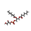  Mass: 442.438 Da / Num. of mol.: 2 / Source method: obtained synthetically / Formula: C18H35O10P Mass: 442.438 Da / Num. of mol.: 2 / Source method: obtained synthetically / Formula: C18H35O10P#5: Chemical | ChemComp-GOL / | Glycerol  Mass: 92.094 Da / Num. of mol.: 1 / Source method: obtained synthetically / Formula: C3H8O3 Mass: 92.094 Da / Num. of mol.: 1 / Source method: obtained synthetically / Formula: C3H8O3Nonpolymer details | FIVE UNKNOWN LIGANDS (UNL) HAVE BEEN MODELED. BASED ON ELECTRON DENSITY AND INTERACTION ...FIVE UNKNOWN LIGANDS (UNL) HAVE BEEN MODELED. BASED ON ELECTRON DENSITY AND INTERACTIO | |
|---|
-Experimental details
-Experiment
| Experiment | Method: X-RAY DIFFRACTION |
|---|
- Sample preparation
| Crystal | Density Matthews: 2.79 Å3/Da / Density % sol: 55.6 % |
|---|---|
| Crystal grow | Temperature: 293 K / Method: vapor diffusion / pH: 6 Details: DROP CONTAINS 10 MG/ML 4E10 FAB, 10 MM PHOSPHATIDYLGLYCEROL (06:0 PG) MIXED 1:1 V/V WITH RESERVOIR SOLUTION OF 0.2 M SODIUM IODIDE, 20% PEG 3350, PH 6.0, VAPOR DIFFUSION, TEMPERATURE 293K |
-Data collection
| Diffraction | Mean temperature: 110 K |
|---|---|
| Diffraction source | Source: SYNCHROTRON / Site: SSRL / Beamline: BL12-2 / Wavelength: 1 Å |
| Detector | Type: DECTRIS PILATUS 6M / Detector: PIXEL / Date: Jun 1, 2013 |
| Radiation | Monochromator: Liquid nitrogen-cooled double crystal, non fixed exit slit Protocol: SINGLE WAVELENGTH / Monochromatic (M) / Laue (L): M / Scattering type: x-ray |
| Radiation wavelength | Wavelength: 1 Å / Relative weight: 1 |
| Reflection | Resolution: 3.96→48.907 Å / Num. obs: 27268 / % possible obs: 95.5 % / Redundancy: 4.43 % / Biso Wilson estimate: 62.9 Å2 / Rsym value: 0.303 / Net I/σ(I): 5.2 |
| Reflection shell | Resolution: 3.96→4.06 Å / Redundancy: 4.16 % / Rmerge(I) obs: 0.711 / Mean I/σ(I) obs: 2.1 / % possible all: 97.4 |
- Processing
| Software |
| |||||||||||||||||||||||||||||||||||||||||||||||||||||||||||||||||||||||||||||
|---|---|---|---|---|---|---|---|---|---|---|---|---|---|---|---|---|---|---|---|---|---|---|---|---|---|---|---|---|---|---|---|---|---|---|---|---|---|---|---|---|---|---|---|---|---|---|---|---|---|---|---|---|---|---|---|---|---|---|---|---|---|---|---|---|---|---|---|---|---|---|---|---|---|---|---|---|---|---|
| Refinement | Method to determine structure: MOLECULAR REPLACEMENT Starting model: PDB ENTRY 2FX7 Resolution: 3.96→48.907 Å / SU ML: 1.19 / Cross valid method: FREE R-VALUE / σ(F): 1.34 / Phase error: 26.8 / Stereochemistry target values: ML
| |||||||||||||||||||||||||||||||||||||||||||||||||||||||||||||||||||||||||||||
| Solvent computation | Shrinkage radii: 0.86 Å / VDW probe radii: 1.1 Å / Solvent model: FLAT BULK SOLVENT MODEL / Bsol: 24.77 Å2 / ksol: 0.31 e/Å3 | |||||||||||||||||||||||||||||||||||||||||||||||||||||||||||||||||||||||||||||
| Displacement parameters | Biso mean: 77.8 Å2
| |||||||||||||||||||||||||||||||||||||||||||||||||||||||||||||||||||||||||||||
| Refinement step | Cycle: LAST / Resolution: 3.96→48.907 Å
| |||||||||||||||||||||||||||||||||||||||||||||||||||||||||||||||||||||||||||||
| Refine LS restraints |
| |||||||||||||||||||||||||||||||||||||||||||||||||||||||||||||||||||||||||||||
| LS refinement shell |
|
