 Movie
Movie Controller


+ Open data

- Basic information
| Entry | Database: PDB / ID: 4xcx | ||||||
|---|---|---|---|---|---|---|---|
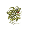
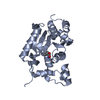
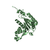
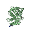
| Title | METHYLTRANSFERASE DOMAIN OF SMALL RNA 2'-O-METHYLTRANSFERASE | ||||||
 Components Components | Small RNA 2'-O-methyltransferase | ||||||
 Keywords Keywords |  TRANSFERASE / METHYLTRANSFERASE / SAH / STRUCTURAL GENOMICS CONSORTIUM / SGC TRANSFERASE / METHYLTRANSFERASE / SAH / STRUCTURAL GENOMICS CONSORTIUM / SGC | ||||||
| Function / homology |  Function and homology information Function and homology informationsmall RNA 2'-O-methyltransferase activity / small RNA 2'-O-methyltransferase / piRNA processing / RNA methyltransferase activity / RNA methylation / siRNA processing / S-adenosylmethionine-dependent methyltransferase activity / O-methyltransferase activity / P granule / PIWI-interacting RNA (piRNA) biogenesis ...small RNA 2'-O-methyltransferase activity / small RNA 2'-O-methyltransferase / piRNA processing / RNA methyltransferase activity / RNA methylation / siRNA processing / S-adenosylmethionine-dependent methyltransferase activity / O-methyltransferase activity / P granule / PIWI-interacting RNA (piRNA) biogenesis / RNA binding / metal ion binding / nucleus / cytoplasmSimilarity search - Function | ||||||
| Biological species |  Homo sapiens (human) Homo sapiens (human) | ||||||
| Method | X-RAY DIFFRACTION / SYNCHROTRON / MOLECULAR REPLACEMENT / Resolution: 2.84 Å | ||||||
 Authors Authors | Walker, J.R. / Zeng, H. / Dong, A. / Li, Y. / Wernimont, A. / Bountra, C. / Arrowsmith, C.H. / Edwards, A.M. / Brown, P.J. / Wu, H. / Structural Genomics Consortium (SGC) | ||||||
 Citation Citation | Journal: To be published Title: Crystal structure of human C1ORF59 in complex with SAH Authors: Walker, J.R. / Zeng, H. / Dong, A. / Li, Y. / Wernimont, A. / Bountra, C. / Arrowsmith, C.H. / Edwards, A.M. / Brown, P.J. / Wu, H. / Structural Genomics Consortium (SGC) | ||||||
| History |
|
- Structure visualization
| Structure viewer | Molecule: MolmilJmol/JSmol |
|---|
- Downloads & links
-Download
| PDBx/mmCIF format | 4xcx.cif.gz | 106.7 KB | Display | PDBx/mmCIF format |
|---|---|---|---|---|
| PDB format | pdb4xcx.ent.gz | 79.2 KB | Display | PDB format |
| PDBx/mmJSON format | 4xcx.json.gz | Tree view | PDBx/mmJSON format | |
| Others |  Other downloads Other downloads |
-Validation report
| Arichive directory | https://data.pdbj.org/pub/pdb/validation_reports/xc/4xcxftp://data.pdbj.org/pub/pdb/validation_reports/xc/4xcx | HTTPS FTP |
|---|
-Related structure data
| Related structure data |  3jwgS S: Starting model for refinement |
|---|---|
| Similar structure data |


-Links
 PDBj
PDBj- Assembly
| Deposited unit | 
| ||||||||
|---|---|---|---|---|---|---|---|---|---|
| 1 |
| ||||||||
| Unit cell |
| ||||||||
| Details | biological unit is the same as asym. |
-Components
| #1: Protein | Mass: 28347.062 Da / Num. of mol.: 1 / Fragment: UNP RESIDUES 14-262 Source method: isolated from a genetically manipulated source Source: (gene. exp.) Homo sapiens (human) / Gene: HENMT1, C1orf59 / Plasmid: PET28-MHL / Production host:  Escherichia coli (E. coli) / Strain (production host): BL21(DE3) Codon Plus RIL Escherichia coli (E. coli) / Strain (production host): BL21(DE3) Codon Plus RILReferences: UniProt: Q5T8I9, Transferases; Transferring one-carbon groups; Methyltransferases |
|---|---|
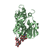
| #2: Chemical | ChemComp-SAH / S-Adenosyl-L-homocysteine  Type: L-peptide linking / Mass: 384.411 Da / Num. of mol.: 1 / Source method: obtained synthetically / Formula: C14H20N6O5S Type: L-peptide linking / Mass: 384.411 Da / Num. of mol.: 1 / Source method: obtained synthetically / Formula: C14H20N6O5S |
| #3: Water | ChemComp-HOH / Water Mass: 18.015 Da / Num. of mol.: 17 / Source method: isolated from a natural source / Formula: H2O Mass: 18.015 Da / Num. of mol.: 17 / Source method: isolated from a natural source / Formula: H2O |
-Experimental details
-Experiment
| Experiment | Method: X-RAY DIFFRACTION / Number of used crystals: 1 |
|---|
- Sample preparation
| Crystal | Density Matthews: 5.21 Å3/Da / Density % sol: 76.38 % |
|---|---|
| Crystal grow | Temperature: 293 K / Method: vapor diffusion, sitting drop / pH: 8.6 Details: 2.4 M NACL, 0.1 M TRIS-HCL, PH 8.6,4% 1,3-PROPANEDIOL |
-Data collection
| Diffraction | Mean temperature: 100 K | ||||||||||||||||||||||||||||||||||||||||||||||||||||||||||||||||||||||||||||||||||||||||||||||||||||||||||||||
|---|---|---|---|---|---|---|---|---|---|---|---|---|---|---|---|---|---|---|---|---|---|---|---|---|---|---|---|---|---|---|---|---|---|---|---|---|---|---|---|---|---|---|---|---|---|---|---|---|---|---|---|---|---|---|---|---|---|---|---|---|---|---|---|---|---|---|---|---|---|---|---|---|---|---|---|---|---|---|---|---|---|---|---|---|---|---|---|---|---|---|---|---|---|---|---|---|---|---|---|---|---|---|---|---|---|---|---|---|---|---|---|
| Diffraction source | Source: SYNCHROTRON / Site: APS  / Beamline: 19-ID / Wavelength: 0.97918 Å / Beamline: 19-ID / Wavelength: 0.97918 Å | ||||||||||||||||||||||||||||||||||||||||||||||||||||||||||||||||||||||||||||||||||||||||||||||||||||||||||||||
| Detector | Type: ADSC QUANTUM 315r / Detector: CCD / Date: Jul 7, 2014 | ||||||||||||||||||||||||||||||||||||||||||||||||||||||||||||||||||||||||||||||||||||||||||||||||||||||||||||||
| Radiation | Protocol: SINGLE WAVELENGTH / Monochromatic (M) / Laue (L): M / Scattering type: x-ray | ||||||||||||||||||||||||||||||||||||||||||||||||||||||||||||||||||||||||||||||||||||||||||||||||||||||||||||||
| Radiation wavelength | Wavelength: 0.97918 Å / Relative weight: 1 | ||||||||||||||||||||||||||||||||||||||||||||||||||||||||||||||||||||||||||||||||||||||||||||||||||||||||||||||
| Reflection | Resolution: 2.84→35 Å / Num. obs: 14604 / % possible obs: 98.4 % / Redundancy: 12.3 % / Biso Wilson estimate: 80.33 Å2 / Rmerge(I) obs: 0.098 / Rpim(I) all: 0.028 / Rrim(I) all: 0.103 / Χ2: 1.095 / Net I/av σ(I): 24.25 / Net I/σ(I): 24.25 / Num. measured all: 180091 | ||||||||||||||||||||||||||||||||||||||||||||||||||||||||||||||||||||||||||||||||||||||||||||||||||||||||||||||
| Reflection shell | Diffraction-ID: 1 / Rejects: 0
|
- Processing
| Software |
| ||||||||||||||||||||||||||||||||||||||||||||||||||||||||||||||||||||||||||||||||||||||||||||||||||||||||||||
|---|---|---|---|---|---|---|---|---|---|---|---|---|---|---|---|---|---|---|---|---|---|---|---|---|---|---|---|---|---|---|---|---|---|---|---|---|---|---|---|---|---|---|---|---|---|---|---|---|---|---|---|---|---|---|---|---|---|---|---|---|---|---|---|---|---|---|---|---|---|---|---|---|---|---|---|---|---|---|---|---|---|---|---|---|---|---|---|---|---|---|---|---|---|---|---|---|---|---|---|---|---|---|---|---|---|---|---|---|---|
| Refinement | Method to determine structure: MOLECULAR REPLACEMENT Starting model: 3JWG Resolution: 2.84→33.98 Å / Cor.coef. Fo:Fc: 0.8825 / Cor.coef. Fo:Fc free: 0.8796 / SU R Cruickshank DPI: 0.726 / Cross valid method: THROUGHOUT / σ(F): 0 / SU R Blow DPI: 0.306 / SU Rfree Blow DPI: 0.243 / SU Rfree Cruickshank DPI: 0.245
| ||||||||||||||||||||||||||||||||||||||||||||||||||||||||||||||||||||||||||||||||||||||||||||||||||||||||||||
| Displacement parameters | Biso max: 186.82 Å2 / Biso mean: 93.03 Å2 / Biso min: 57.87 Å2
| ||||||||||||||||||||||||||||||||||||||||||||||||||||||||||||||||||||||||||||||||||||||||||||||||||||||||||||
| Refine analyze | Luzzati coordinate error obs: 0.575 Å | ||||||||||||||||||||||||||||||||||||||||||||||||||||||||||||||||||||||||||||||||||||||||||||||||||||||||||||
| Refinement step | Cycle: final / Resolution: 2.84→33.98 Å
| ||||||||||||||||||||||||||||||||||||||||||||||||||||||||||||||||||||||||||||||||||||||||||||||||||||||||||||
| Refine LS restraints |
| ||||||||||||||||||||||||||||||||||||||||||||||||||||||||||||||||||||||||||||||||||||||||||||||||||||||||||||
| LS refinement shell | Resolution: 2.84→3.07 Å / Total num. of bins used: 7
| ||||||||||||||||||||||||||||||||||||||||||||||||||||||||||||||||||||||||||||||||||||||||||||||||||||||||||||
| Refinement TLS params. | Method: refined / Origin x: 33.9105 Å / Origin y: 98.0601 Å / Origin z: 9.4459 Å
| ||||||||||||||||||||||||||||||||||||||||||||||||||||||||||||||||||||||||||||||||||||||||||||||||||||||||||||
| Refinement TLS group | Selection details: { A|26 - A|400 } |
