 Movie
Movie Controller


+ Open data

- Basic information
| Entry | Database: PDB / ID: 4xb0 | |||||||||
|---|---|---|---|---|---|---|---|---|---|---|
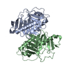
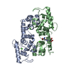
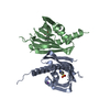
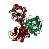
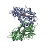
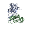
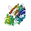
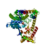
| Title | Structure of the Plk2 polo-box domain | |||||||||
 Components Components | Serine/threonine-protein kinase PLK2 | |||||||||
 Keywords Keywords |  TRANSFERASE / polo-box domain / Polo-like kinase 2 / Plk2 TRANSFERASE / polo-box domain / Polo-like kinase 2 / Plk2 | |||||||||
| Function / homology |  Function and homology information Function and homology informationregulation of centriole replication / Rap protein signal transduction / negative regulation of apoptotic process in bone marrow cell / polo kinase / ATP-dependent protein binding / long-term synaptic depression / TP53 regulates transcription of additional cell cycle genes whose exact role in the p53 pathway remain uncertain / CD163 mediating an anti-inflammatory response / positive regulation of cell migration involved in sprouting angiogenesis / negative regulation of cellular senescence ...regulation of centriole replication / Rap protein signal transduction / negative regulation of apoptotic process in bone marrow cell / polo kinase / ATP-dependent protein binding / long-term synaptic depression / TP53 regulates transcription of additional cell cycle genes whose exact role in the p53 pathway remain uncertain / CD163 mediating an anti-inflammatory response / positive regulation of cell migration involved in sprouting angiogenesis / negative regulation of cellular senescence / positive regulation of autophagy / negative regulation of inflammatory response to antigenic stimulus / DNA damage response, signal transduction by p53 class mediator resulting in cell cycle arrest / NPAS4 regulates expression of target genes / centriole / negative regulation of angiogenesis / mitotic spindle organization / long-term synaptic potentiation / regulation of synaptic plasticity / G1/S transition of mitotic cell cycle / kinetochore / memory / spindle pole / positive regulation of protein catabolic process / peptidyl-serine phosphorylation / positive regulation of canonical NF-kappaB signal transduction / Ras protein signal transduction / protein phosphorylation / protein serine kinase activity / protein serine/threonine kinase activity / centrosome / dendrite / chromatin / negative regulation of apoptotic process / ATP binding / nucleus / cytosol / cytoplasmSimilarity search - Function | |||||||||
| Biological species |  Homo sapiens (human) Homo sapiens (human) | |||||||||
| Method | X-RAY DIFFRACTION / SYNCHROTRON / MOLECULAR REPLACEMENT / Resolution: 2.701 Å | |||||||||
 Authors Authors | Kim, J.H. / Ku, B. / Kim, S.J. | |||||||||
| Funding support |  Korea, Republic Of, 2items Korea, Republic Of, 2items
| |||||||||
 Citation Citation | Journal: Proteins / Year: 2015 Title: Structural analysis of the polo-box domain of human Polo-like kinase 2 Authors: Kim, J.H. / Ku, B. / Lee, K.S. / Kim, S.J. | |||||||||
| History |
|
- Structure visualization
| Structure viewer | Molecule: MolmilJmol/JSmol |
|---|
- Downloads & links
-Download
| PDBx/mmCIF format | 4xb0.cif.gz | 97.8 KB | Display | PDBx/mmCIF format |
|---|---|---|---|---|
| PDB format | pdb4xb0.ent.gz | 75.1 KB | Display | PDB format |
| PDBx/mmJSON format | 4xb0.json.gz | Tree view | PDBx/mmJSON format | |
| Others |  Other downloads Other downloads |
-Validation report
| Arichive directory | https://data.pdbj.org/pub/pdb/validation_reports/xb/4xb0ftp://data.pdbj.org/pub/pdb/validation_reports/xb/4xb0 | HTTPS FTP |
|---|
-Related structure data
| Similar structure data |
|---|


-Links
 PDBj
PDBj


- Assembly
| Deposited unit | 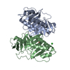
| ||||||||
|---|---|---|---|---|---|---|---|---|---|
| 1 |
| ||||||||
| Unit cell |
|
-Components
| #1: Protein | Mass: 25161.703 Da / Num. of mol.: 2 / Fragment: polo-box domain, UNP residues 468-685 Source method: isolated from a genetically manipulated source Source: (gene. exp.) Homo sapiens (human) / Gene: PLK2, SNK / Production host:  Escherichia coli (E. coli) / Strain (production host): BL21(DE3) RIL / References: UniProt: Q9NYY3, polo kinase Escherichia coli (E. coli) / Strain (production host): BL21(DE3) RIL / References: UniProt: Q9NYY3, polo kinase#2: Chemical | ChemComp-PO4 / | Phosphate  Mass: 94.971 Da / Num. of mol.: 1 / Source method: obtained synthetically / Formula: PO4 Mass: 94.971 Da / Num. of mol.: 1 / Source method: obtained synthetically / Formula: PO4#3: Chemical | Chloride  Mass: 35.453 Da / Num. of mol.: 2 / Source method: obtained synthetically / Formula: Cl Mass: 35.453 Da / Num. of mol.: 2 / Source method: obtained synthetically / Formula: Cl#4: Water | ChemComp-HOH / | Water Mass: 18.015 Da / Num. of mol.: 22 / Source method: isolated from a natural source / Formula: H2O Mass: 18.015 Da / Num. of mol.: 22 / Source method: isolated from a natural source / Formula: H2O |
|---|
-Experimental details
-Experiment
| Experiment | Method: X-RAY DIFFRACTION |
|---|
- Sample preparation
| Crystal | Density Matthews: 3 Å3/Da / Density % sol: 59.03 % |
|---|---|
| Crystal grow | Temperature: 291 K / Method: vapor diffusion, sitting drop Details: 1.26 M sodium phosphate, 0.14 M potassium phosphate |
-Data collection
| Diffraction | Mean temperature: 93 K |
|---|---|
| Diffraction source | Source: SYNCHROTRON / Site: PAL/PLS / Beamline: 7A (6B, 6C1) / Wavelength: 0.97949 Å |
| Detector | Type: ADSC QUANTUM 210 / Detector: CCD / Date: Oct 2, 2014 |
| Radiation | Protocol: SINGLE WAVELENGTH / Monochromatic (M) / Laue (L): M / Scattering type: x-ray |
| Radiation wavelength | Wavelength: 0.97949 Å / Relative weight: 1 |
| Reflection | Resolution: 2.7→50 Å / Num. obs: 16508 / % possible obs: 99.6 % / Redundancy: 22.1 % / Net I/σ(I): 48.1 |
- Processing
| Software |
| |||||||||||||||||||||||||||||||||||||||||||||||||||||||||||||||||||||||||||||||||||||||||||
|---|---|---|---|---|---|---|---|---|---|---|---|---|---|---|---|---|---|---|---|---|---|---|---|---|---|---|---|---|---|---|---|---|---|---|---|---|---|---|---|---|---|---|---|---|---|---|---|---|---|---|---|---|---|---|---|---|---|---|---|---|---|---|---|---|---|---|---|---|---|---|---|---|---|---|---|---|---|---|---|---|---|---|---|---|---|---|---|---|---|---|---|---|
| Refinement | Method to determine structure: MOLECULAR REPLACEMENT Starting model: the polo-box domain of Plk1 Resolution: 2.701→26.922 Å / SU ML: 0.4 / Cross valid method: NONE / σ(F): 1.76 / Phase error: 26.18 / Stereochemistry target values: ML
| |||||||||||||||||||||||||||||||||||||||||||||||||||||||||||||||||||||||||||||||||||||||||||
| Solvent computation | Shrinkage radii: 0.9 Å / VDW probe radii: 1.11 Å / Solvent model: FLAT BULK SOLVENT MODEL | |||||||||||||||||||||||||||||||||||||||||||||||||||||||||||||||||||||||||||||||||||||||||||
| Refinement step | Cycle: LAST / Resolution: 2.701→26.922 Å
| |||||||||||||||||||||||||||||||||||||||||||||||||||||||||||||||||||||||||||||||||||||||||||
| Refine LS restraints |
| |||||||||||||||||||||||||||||||||||||||||||||||||||||||||||||||||||||||||||||||||||||||||||
| LS refinement shell |
|
