 Movie
Movie Controller


+ Open data

- Basic information
| Entry | Database: PDB / ID: 4x41 | ||||||
|---|---|---|---|---|---|---|---|
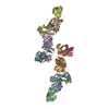
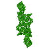
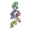
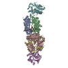
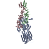
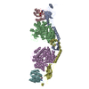
| Title | Crystal Structure of Protein Arginine Methyltransferase PRMT8 | ||||||
 Components Components | Protein arginine N-methyltransferase 8 | ||||||
 Keywords Keywords |  TRANSFERASE / protein arginine methyltransferase / methylation / dimerization TRANSFERASE / protein arginine methyltransferase / methylation / dimerization | ||||||
| Function / homology |  Function and homology information Function and homology informationprotein-arginine omega-N monomethyltransferase activity / histone H4 methyltransferase activity / peptidyl-arginine methylation / type I protein arginine methyltransferase / protein-arginine omega-N asymmetric methyltransferase activity / regulation of protein binding / protein methylation / S-adenosylmethionine-dependent methyltransferase activity / S-adenosyl-L-methionine binding / protein homooligomerization ...protein-arginine omega-N monomethyltransferase activity / histone H4 methyltransferase activity / peptidyl-arginine methylation / type I protein arginine methyltransferase / protein-arginine omega-N asymmetric methyltransferase activity / regulation of protein binding / protein methylation / S-adenosylmethionine-dependent methyltransferase activity / S-adenosyl-L-methionine binding / protein homooligomerization / cytoplasmic side of plasma membrane / enzyme binding / protein homodimerization activity / identical protein binding / plasma membraneSimilarity search - Function | ||||||
| Biological species |  Homo sapiens (human) Homo sapiens (human) | ||||||
| Method | X-RAY DIFFRACTION / SYNCHROTRON / MOLECULAR REPLACEMENT / Resolution: 3.5 Å | ||||||
 Authors Authors | Lee, W.C. / Ho, M.C. | ||||||
 Citation Citation | Journal: Biochemistry / Year: 2015 Title: Protein Arginine Methyltransferase 8: Tetrameric Structure and Protein Substrate Specificity Authors: Lee, W.C. / Lin, W.L. / Matsui, T. / Chen, E.S. / Wei, T.Y. / Lin, W.H. / Hu, H. / Zheng, Y.G. / Tsai, M.D. / Ho, M.C. | ||||||
| History |
|
- Structure visualization
| Structure viewer | Molecule: MolmilJmol/JSmol |
|---|
- Downloads & links
-Download
| PDBx/mmCIF format | 4x41.cif.gz | 137.4 KB | Display | PDBx/mmCIF format |
|---|---|---|---|---|
| PDB format | pdb4x41.ent.gz | 105.8 KB | Display | PDB format |
| PDBx/mmJSON format | 4x41.json.gz | Tree view | PDBx/mmJSON format | |
| Others |  Other downloads Other downloads |
-Validation report
| Arichive directory | https://data.pdbj.org/pub/pdb/validation_reports/x4/4x41ftp://data.pdbj.org/pub/pdb/validation_reports/x4/4x41 | HTTPS FTP |
|---|
-Related structure data
| Similar structure data |
|---|


-Links
 PDBj
PDBj- Assembly
| Deposited unit | 
| ||||||||
|---|---|---|---|---|---|---|---|---|---|
| 1 | 
| ||||||||
| Unit cell |
|
-Components
| #1: Protein | Mass: 41614.520 Da / Num. of mol.: 2 / Fragment: UNP residues 52-385 Source method: isolated from a genetically manipulated source Source: (gene. exp.) Homo sapiens (human) / Gene: PRMT8, HRMT1L3, HRMT1L4 / Production host:  Escherichia coli (E. coli) / References: UniProt: Q9NR22, EC: 2.1.1.125 Escherichia coli (E. coli) / References: UniProt: Q9NR22, EC: 2.1.1.125#2: Chemical | ChemComp-SAH / | S-Adenosyl-L-homocysteine  Type: L-peptide linking / Mass: 384.411 Da / Num. of mol.: 1 / Source method: obtained synthetically / Formula: C14H20N6O5S Type: L-peptide linking / Mass: 384.411 Da / Num. of mol.: 1 / Source method: obtained synthetically / Formula: C14H20N6O5SSequence details | THIS SEQUENCE IS BASED ON GENBANK AAH22458.2. RESIDUE 141 IS GLU IN AAH22458.2. | |
|---|
-Experimental details
-Experiment
| Experiment | Method: X-RAY DIFFRACTION / Number of used crystals: 1 |
|---|
- Sample preparation
| Crystal | Density Matthews: 3.27 Å3/Da / Density % sol: 67.38 % |
|---|---|
| Crystal grow | Temperature: 298 K / Method: vapor diffusion, sitting drop / pH: 7.5 Details: 150mM D-glucose, 100mM HEPES/MOPS pH7.5, 40% Glycerol/PEG4000 |
-Data collection
| Diffraction | Mean temperature: 100 K | |||||||||||||||||||||||||||||||||||||||||||||||||||||||||||||||||||||||||||||||||||||||||||||||||||
|---|---|---|---|---|---|---|---|---|---|---|---|---|---|---|---|---|---|---|---|---|---|---|---|---|---|---|---|---|---|---|---|---|---|---|---|---|---|---|---|---|---|---|---|---|---|---|---|---|---|---|---|---|---|---|---|---|---|---|---|---|---|---|---|---|---|---|---|---|---|---|---|---|---|---|---|---|---|---|---|---|---|---|---|---|---|---|---|---|---|---|---|---|---|---|---|---|---|---|---|---|
| Diffraction source | Source: SYNCHROTRON / Site: NSRRC  / Beamline: BL13B1 / Wavelength: 1 Å / Beamline: BL13B1 / Wavelength: 1 Å | |||||||||||||||||||||||||||||||||||||||||||||||||||||||||||||||||||||||||||||||||||||||||||||||||||
| Detector | Type: RAYONIX MX-300 / Detector: CCD / Date: May 22, 2014 | |||||||||||||||||||||||||||||||||||||||||||||||||||||||||||||||||||||||||||||||||||||||||||||||||||
| Radiation | Protocol: SINGLE WAVELENGTH / Monochromatic (M) / Laue (L): M / Scattering type: x-ray | |||||||||||||||||||||||||||||||||||||||||||||||||||||||||||||||||||||||||||||||||||||||||||||||||||
| Radiation wavelength | Wavelength: 1 Å / Relative weight: 1 | |||||||||||||||||||||||||||||||||||||||||||||||||||||||||||||||||||||||||||||||||||||||||||||||||||
| Reflection | Resolution: 3.5→20 Å / Num. obs: 13561 / % possible obs: 94.7 % / Redundancy: 2.9 % / Biso Wilson estimate: 58.37 Å2 / Rmerge(I) obs: 0.196 / Rpim(I) all: 0.133 / Rrim(I) all: 0.239 / Χ2: 0.995 / Net I/av σ(I): 3.979 / Net I/σ(I): 5.7 / Num. measured all: 38801 | |||||||||||||||||||||||||||||||||||||||||||||||||||||||||||||||||||||||||||||||||||||||||||||||||||
| Reflection shell | Diffraction-ID: 1 / Rejects: 0
|
- Processing
| Software |
| ||||||||||||||||||||||||||||||||||||||||||
|---|---|---|---|---|---|---|---|---|---|---|---|---|---|---|---|---|---|---|---|---|---|---|---|---|---|---|---|---|---|---|---|---|---|---|---|---|---|---|---|---|---|---|---|
| Refinement | Method to determine structure: MOLECULAR REPLACEMENT / Resolution: 3.5→19.727 Å / SU ML: 0.44 / Cross valid method: FREE R-VALUE / σ(F): 1.33 / Phase error: 28.28 / Stereochemistry target values: ML
| ||||||||||||||||||||||||||||||||||||||||||
| Solvent computation | Shrinkage radii: 0.9 Å / VDW probe radii: 1.11 Å / Solvent model: FLAT BULK SOLVENT MODEL | ||||||||||||||||||||||||||||||||||||||||||
| Displacement parameters | Biso max: 157.04 Å2 / Biso mean: 55.274 Å2 / Biso min: 20.35 Å2 | ||||||||||||||||||||||||||||||||||||||||||
| Refinement step | Cycle: final / Resolution: 3.5→19.727 Å
| ||||||||||||||||||||||||||||||||||||||||||
| Refine LS restraints |
| ||||||||||||||||||||||||||||||||||||||||||
| LS refinement shell | Refine-ID: X-RAY DIFFRACTION / Total num. of bins used: 5
|
