 Movie
Movie Controller


+ Open data

- Basic information
| Entry | Database: PDB / ID: 4x0j | ||||||
|---|---|---|---|---|---|---|---|
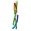
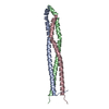
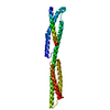
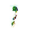
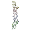
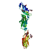
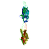
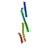
| Title | Trypanosoma brucei haptoglobin-haemoglobin receptor | ||||||
 Components Components | Haptoglobin-hemoglobin receptor | ||||||
 Keywords Keywords |  PROTEIN TRANSPORT / Trypanosoma brucei / Haptoglobin-haemoglobin receptor PROTEIN TRANSPORT / Trypanosoma brucei / Haptoglobin-haemoglobin receptor | ||||||
| Function / homology | Ferritin - #80 / Ferritin / Up-down Bundle / Mainly Alpha / Haptoglobin-hemoglobin receptor Function and homology information Function and homology information | ||||||
| Biological species |  Trypanosoma brucei brucei (eukaryote) Trypanosoma brucei brucei (eukaryote) | ||||||
| Method | X-RAY DIFFRACTION / SYNCHROTRON / MOLECULAR REPLACEMENT / Resolution: 1.85 Å | ||||||
 Authors Authors | Lane-Serff, H. / MacGregor, P. / Lowe, E.D. / Carrington, M. / Higgins, M.K. | ||||||
 Citation Citation | Journal: Elife / Year: 2014 Title: Structural basis for ligand and innate immunity factor uptake by the trypanosome haptoglobin-haemoglobin receptor. Authors: Lane-Serff, H. / MacGregor, P. / Lowe, E.D. / Carrington, M. / Higgins, M.K. | ||||||
| History |
|
- Structure visualization
| Structure viewer | Molecule: MolmilJmol/JSmol |
|---|
- Downloads & links
-Download
| PDBx/mmCIF format | 4x0j.cif.gz | 114.6 KB | Display | PDBx/mmCIF format |
|---|---|---|---|---|
| PDB format | pdb4x0j.ent.gz | 88.3 KB | Display | PDB format |
| PDBx/mmJSON format | 4x0j.json.gz | Tree view | PDBx/mmJSON format | |
| Others |  Other downloads Other downloads |
-Validation report
| Arichive directory | https://data.pdbj.org/pub/pdb/validation_reports/x0/4x0jftp://data.pdbj.org/pub/pdb/validation_reports/x0/4x0j | HTTPS FTP |
|---|
-Related structure data
| Related structure data |  4x0lC 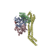 5hu6C  4x0i S: Starting model for refinement C: citing same article ( |
|---|---|
| Similar structure data |


-Links
 PDBj
PDBj- Assembly
| Deposited unit | 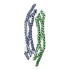
| ||||||||||||||||||
|---|---|---|---|---|---|---|---|---|---|---|---|---|---|---|---|---|---|---|---|
| 1 | 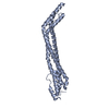
| ||||||||||||||||||
| 2 | 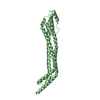
| ||||||||||||||||||
| Unit cell |
| ||||||||||||||||||
| Noncrystallographic symmetry (NCS) | NCS domain:
NCS domain segments: Component-ID: 0 / Ens-ID: 1 / Beg auth comp-ID: GLU / Beg label comp-ID: GLU / End auth comp-ID: GLU / End label comp-ID: GLU / Refine code: 0 / Auth seq-ID: 37 - 296 / Label seq-ID: 3 - 262
|
-Components
| #1: Protein | Mass: 28502.127 Da / Num. of mol.: 2 Source method: isolated from a genetically manipulated source Source: (gene. exp.) Trypanosoma brucei brucei (eukaryote) / Gene: HpHbR / Production host:  Escherichia coli (E. coli) / References: UniProt: I7BA99 Escherichia coli (E. coli) / References: UniProt: I7BA99#2: Water | ChemComp-HOH / | Water Mass: 18.015 Da / Num. of mol.: 264 / Source method: isolated from a natural source / Formula: H2O Mass: 18.015 Da / Num. of mol.: 264 / Source method: isolated from a natural source / Formula: H2O |
|---|
-Experimental details
-Experiment
| Experiment | Method: X-RAY DIFFRACTION |
|---|
- Sample preparation
| Crystal | Density Matthews: 2.38 Å3/Da / Density % sol: 48.23 % |
|---|---|
| Crystal grow | Temperature: 291 K / Method: vapor diffusion, sitting drop / Details: 0.15 M KBr, 30 % w/v PEG 2000 MME |
-Data collection
| Diffraction | Mean temperature: 100 K |
|---|---|
| Diffraction source | Source: SYNCHROTRON / Site: Diamond  / Beamline: I03 / Wavelength: 0.9763 Å / Beamline: I03 / Wavelength: 0.9763 Å |
| Detector | Type: DECTRIS PILATUS 6M / Detector: PIXEL / Date: Feb 24, 2013 |
| Radiation | Protocol: SINGLE WAVELENGTH / Monochromatic (M) / Laue (L): M / Scattering type: x-ray |
| Radiation wavelength | Wavelength: 0.9763 Å / Relative weight: 1 |
| Reflection | Resolution: 1.85→203.14 Å / Num. obs: 44672 / % possible obs: 97.4 % / Redundancy: 3.1 % / Rmerge(I) obs: 0.099 / Net I/σ(I): 10.2 |
| Reflection shell | Resolution: 1.85→1.95 Å / Redundancy: 3.1 % / Rmerge(I) obs: 0.64 / % possible all: 96.4 |
- Processing
| Software |
| ||||||||||||||||||||||||||||||||||||||||||||||||||||||||||||||||||||||||||||||||||||||||||||||||||||||||||||||||||||||||||||||||||||||||||||||||||||||||||||||||||||||||||||||||||||||
|---|---|---|---|---|---|---|---|---|---|---|---|---|---|---|---|---|---|---|---|---|---|---|---|---|---|---|---|---|---|---|---|---|---|---|---|---|---|---|---|---|---|---|---|---|---|---|---|---|---|---|---|---|---|---|---|---|---|---|---|---|---|---|---|---|---|---|---|---|---|---|---|---|---|---|---|---|---|---|---|---|---|---|---|---|---|---|---|---|---|---|---|---|---|---|---|---|---|---|---|---|---|---|---|---|---|---|---|---|---|---|---|---|---|---|---|---|---|---|---|---|---|---|---|---|---|---|---|---|---|---|---|---|---|---|---|---|---|---|---|---|---|---|---|---|---|---|---|---|---|---|---|---|---|---|---|---|---|---|---|---|---|---|---|---|---|---|---|---|---|---|---|---|---|---|---|---|---|---|---|---|---|---|---|
| Refinement | Method to determine structure: MOLECULAR REPLACEMENT Starting model: 4X0I 4x0i Resolution: 1.85→203.14 Å / Cor.coef. Fo:Fc: 0.953 / Cor.coef. Fo:Fc free: 0.932 / SU B: 3.942 / SU ML: 0.114 / Cross valid method: THROUGHOUT / ESU R: 0.153 / ESU R Free: 0.144 / Stereochemistry target values: MAXIMUM LIKELIHOOD / Details: HYDROGENS HAVE BEEN ADDED IN THE RIDING POSITIONS
| ||||||||||||||||||||||||||||||||||||||||||||||||||||||||||||||||||||||||||||||||||||||||||||||||||||||||||||||||||||||||||||||||||||||||||||||||||||||||||||||||||||||||||||||||||||||
| Solvent computation | Ion probe radii: 0.8 Å / Shrinkage radii: 0.8 Å / VDW probe radii: 1.2 Å / Solvent model: MASK | ||||||||||||||||||||||||||||||||||||||||||||||||||||||||||||||||||||||||||||||||||||||||||||||||||||||||||||||||||||||||||||||||||||||||||||||||||||||||||||||||||||||||||||||||||||||
| Displacement parameters | Biso mean: 30.564 Å2
| ||||||||||||||||||||||||||||||||||||||||||||||||||||||||||||||||||||||||||||||||||||||||||||||||||||||||||||||||||||||||||||||||||||||||||||||||||||||||||||||||||||||||||||||||||||||
| Refinement step | Cycle: 1 / Resolution: 1.85→203.14 Å
| ||||||||||||||||||||||||||||||||||||||||||||||||||||||||||||||||||||||||||||||||||||||||||||||||||||||||||||||||||||||||||||||||||||||||||||||||||||||||||||||||||||||||||||||||||||||
| Refine LS restraints |
|