[English] 日本語
Yorodumi
- PDB-4wtq: Structure of the Ssl1 laccase mutant M295L -

+
Open data


ID or keywords:

Loading...

-
Basic information

Entry
Database: PDB / ID: 4wtq
TitleStructure of the Ssl1 laccase mutant M295L
ComponentsCopper oxidase
KeywordsOXIDOREDUCTASE / Laccase / multicopper oxidase
Function / homology
Function and homology information


oxidoreductase activity / copper ion binding
Similarity search - Function
Multicopper oxidase, copper-binding site / Multicopper oxidases signature 2. / Multicopper oxidase / Multicopper oxidase, C-terminal / Multicopper oxidase / Multicopper oxidase, N-terminal / Multicopper oxidase / Cupredoxins - blue copper proteins / Twin arginine translocation (Tat) signal profile. / Twin-arginine translocation pathway, signal sequence ...Multicopper oxidase, copper-binding site / Multicopper oxidases signature 2. / Multicopper oxidase / Multicopper oxidase, C-terminal / Multicopper oxidase / Multicopper oxidase, N-terminal / Multicopper oxidase / Cupredoxins - blue copper proteins / Twin arginine translocation (Tat) signal profile. / Twin-arginine translocation pathway, signal sequence / Cupredoxin / Immunoglobulin-like / Sandwich / Mainly Beta
Similarity search - Domain/homology
COPPER (II) ION / OXYGEN ATOM / Copper oxidase
Similarity search - Component
Biological speciesStreptomyces sviceus ATCC 29083 (bacteria)
MethodX-RAY DIFFRACTION / SYNCHROTRON / MOLECULAR REPLACEMENT / Resolution: 1.8 Å
AuthorsGunne, M. / Hoeppner, A. / Urlacher, V.B.
CitationJournal: To Be Published
Title: Structure of the Ssl1 laccase mutant M295L
Authors: Gunne, M. / Hoeppner, A. / Urlacher, V.B.
History
DepositionOct 30, 2014Deposition site: RCSB / Processing site: PDBE
Revision 1.0Dec 23, 2015Provider: repository / Type: Initial release
Revision 1.1Mar 7, 2018Group: Data collection / Category: diffrn_source / Item: _diffrn_source.pdbx_synchrotron_site
Revision 1.2Jan 10, 2024Group: Data collection / Database references ...Data collection / Database references / Derived calculations / Refinement description
Category: chem_comp_atom / chem_comp_bond ...chem_comp_atom / chem_comp_bond / database_2 / pdbx_initial_refinement_model / pdbx_struct_conn_angle / struct_conn
Item: _database_2.pdbx_DOI / _database_2.pdbx_database_accession ..._database_2.pdbx_DOI / _database_2.pdbx_database_accession / _pdbx_struct_conn_angle.ptnr1_auth_asym_id / _pdbx_struct_conn_angle.ptnr1_auth_comp_id / _pdbx_struct_conn_angle.ptnr1_auth_seq_id / _pdbx_struct_conn_angle.ptnr1_label_asym_id / _pdbx_struct_conn_angle.ptnr1_label_atom_id / _pdbx_struct_conn_angle.ptnr1_label_comp_id / _pdbx_struct_conn_angle.ptnr1_label_seq_id / _pdbx_struct_conn_angle.ptnr3_auth_asym_id / _pdbx_struct_conn_angle.ptnr3_auth_comp_id / _pdbx_struct_conn_angle.ptnr3_auth_seq_id / _pdbx_struct_conn_angle.ptnr3_label_asym_id / _pdbx_struct_conn_angle.ptnr3_label_atom_id / _pdbx_struct_conn_angle.ptnr3_label_comp_id / _pdbx_struct_conn_angle.ptnr3_label_seq_id / _pdbx_struct_conn_angle.value / _struct_conn.pdbx_dist_value / _struct_conn.ptnr1_auth_asym_id / _struct_conn.ptnr1_auth_comp_id / _struct_conn.ptnr1_auth_seq_id / _struct_conn.ptnr1_label_asym_id / _struct_conn.ptnr1_label_atom_id / _struct_conn.ptnr1_label_comp_id / _struct_conn.ptnr1_label_seq_id / _struct_conn.ptnr2_auth_asym_id / _struct_conn.ptnr2_auth_comp_id / _struct_conn.ptnr2_auth_seq_id / _struct_conn.ptnr2_label_asym_id / _struct_conn.ptnr2_label_atom_id / _struct_conn.ptnr2_label_comp_id / _struct_conn.ptnr2_label_seq_id

-
Structure visualization

Structure viewerMolecule:
MolmilJmol/JSmol

Downloads & links

-
Assembly

Deposited unit
A: Copper oxidase
B: Copper oxidase
C: Copper oxidase
hetero molecules


Theoretical massNumber of molelcules
Total (without water)98,16915
Polymers97,4733
Non-polymers69612
Water7,764431
1


  • Idetical with deposited unit
  • defined by author&software
TypeNameSymmetry operationNumber
identity operation1_555x,y,z1
Buried area11740 Å2
ΔGint-118 kcal/mol
Surface area27300 Å2
MethodPISA
Unit cell
Length a, b, c (Å)51.528, 104.795, 164.389
Angle α, β, γ (deg.)90.00, 90.00, 90.00
Int Tables number19
Space group name H-MP212121

-
Components

#1: Protein Copper oxidase


Mass: 32491.039 Da / Num. of mol.: 3 / Mutation: M295L
Source method: isolated from a genetically manipulated source
Source: (gene. exp.) Streptomyces sviceus ATCC 29083 (bacteria)
Gene: SSEG_02446 / Production host: Escherichia coli (E. coli) / References: UniProt: B5HSR1, laccase
#2: Chemical
ChemComp-CU / COPPER (II) ION / Copper


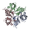
Mass: 63.546 Da / Num. of mol.: 9 / Source method: obtained synthetically / Formula: Cu
#3: Chemical ChemComp-O / OXYGEN ATOM / Oxygen


Mass: 15.999 Da / Num. of mol.: 2 / Source method: obtained synthetically / Formula: O
#4: Chemical ChemComp-GOL / GLYCEROL / GLYCERIN / PROPANE-1,2,3-TRIOL / Glycerol


Mass: 92.094 Da / Num. of mol.: 1 / Source method: obtained synthetically / Formula: C3H8O3
#5: Water ChemComp-HOH / water / Water


Mass: 18.015 Da / Num. of mol.: 431 / Source method: isolated from a natural source / Formula: H2O

-
Experimental details

-
Experiment

ExperimentMethod: X-RAY DIFFRACTION

-
Sample preparation

CrystalDensity Matthews: 2.28 Å3/Da / Density % sol: 45.98 %
Crystal growTemperature: 293 K / Method: vapor diffusion, sitting drop / pH: 7
Details: 50 mM HEPES, pH 7.0, 1.0-1.3 M ammonium sulfate, 5-20 mM hexamminecobalt(III) chloride

-
Data collection

DiffractionMean temperature: 100 K
Diffraction sourceSource: SYNCHROTRON / Site: PETRA III, EMBL c/o DESY / Beamline: P14 (MX2) / Wavelength: 0.900002 Å
DetectorType: DECTRIS PILATUS 6M / Detector: PIXEL / Date: Apr 25, 2013
RadiationProtocol: SINGLE WAVELENGTH / Monochromatic (M) / Laue (L): M / Scattering type: x-ray
Radiation wavelengthWavelength: 0.900002 Å / Relative weight: 1
ReflectionResolution: 1.8→24.96 Å / Num. obs: 82580 / % possible obs: 99.1 % / Redundancy: 6.3 % / Rmerge(I) obs: 0.07 / Net I/σ(I): 15.3
Reflection shellResolution: 1.8→1.9 Å / Redundancy: 6.8 % / Rmerge(I) obs: 0.508 / Mean I/σ(I) obs: 4 / % possible all: 99.3

-
Processing

Software
NameVersionClassification
REFMAC5.8.0071refinement
XDSdata reduction
SCALAdata scaling
PHASERphasing
Cootmodel building
RefinementMethod to determine structure: MOLECULAR REPLACEMENT
Starting model: 4M3H
Resolution: 1.8→24.58 Å / Cor.coef. Fo:Fc: 0.965 / Cor.coef. Fo:Fc free: 0.948 / SU B: 2.823 / SU ML: 0.085 / Cross valid method: THROUGHOUT / ESU R: 0.115 / ESU R Free: 0.113 / Stereochemistry target values: MAXIMUM LIKELIHOOD / Details: HYDROGENS HAVE BEEN ADDED IN THE RIDING POSITIONS
RfactorNum. reflection% reflectionSelection details
Rfree0.20812 4083 5 %RANDOM
Rwork0.17094 ---
obs0.17277 77770 98.11 %-
Solvent computationIon probe radii: 0.8 Å / Shrinkage radii: 0.8 Å / VDW probe radii: 1.2 Å / Solvent model: MASK
Displacement parametersBiso mean: 27.946 Å2
Baniso -1Baniso -2Baniso -3
1-0.01 Å20 Å20 Å2
2---0.01 Å20 Å2
3---0 Å2
Refinement stepCycle: 1 / Resolution: 1.8→24.58 Å
ProteinNucleic acidLigandSolventTotal
Num. atoms6270 0 17 431 6718
Refine LS restraints
Refine-IDTypeDev idealDev ideal targetNumber
X-RAY DIFFRACTIONr_bond_refined_d0.0190.0196449
X-RAY DIFFRACTIONr_bond_other_d0.0020.025868
X-RAY DIFFRACTIONr_angle_refined_deg1.8591.9228750
X-RAY DIFFRACTIONr_angle_other_deg0.852313463
X-RAY DIFFRACTIONr_dihedral_angle_1_deg6.8145805
X-RAY DIFFRACTIONr_dihedral_angle_2_deg31.42422.785316
X-RAY DIFFRACTIONr_dihedral_angle_3_deg14.3115974
X-RAY DIFFRACTIONr_dihedral_angle_4_deg16.5491551
X-RAY DIFFRACTIONr_chiral_restr0.1150.2908
X-RAY DIFFRACTIONr_gen_planes_refined0.010.0217466
X-RAY DIFFRACTIONr_gen_planes_other0.0010.021609
X-RAY DIFFRACTIONr_nbd_refined
X-RAY DIFFRACTIONr_nbd_other
X-RAY DIFFRACTIONr_nbtor_refined
X-RAY DIFFRACTIONr_nbtor_other
X-RAY DIFFRACTIONr_xyhbond_nbd_refined
X-RAY DIFFRACTIONr_xyhbond_nbd_other
X-RAY DIFFRACTIONr_metal_ion_refined
X-RAY DIFFRACTIONr_metal_ion_other
X-RAY DIFFRACTIONr_symmetry_vdw_refined
X-RAY DIFFRACTIONr_symmetry_vdw_other
X-RAY DIFFRACTIONr_symmetry_hbond_refined
X-RAY DIFFRACTIONr_symmetry_hbond_other
X-RAY DIFFRACTIONr_symmetry_metal_ion_refined
X-RAY DIFFRACTIONr_symmetry_metal_ion_other
X-RAY DIFFRACTIONr_mcbond_it2.6672.4953229
X-RAY DIFFRACTIONr_mcbond_other2.6622.4943228
X-RAY DIFFRACTIONr_mcangle_it3.7753.7284031
X-RAY DIFFRACTIONr_mcangle_other3.7753.7294032
X-RAY DIFFRACTIONr_scbond_it3.8352.9233220
X-RAY DIFFRACTIONr_scbond_other3.8352.9233220
X-RAY DIFFRACTIONr_scangle_it
X-RAY DIFFRACTIONr_scangle_other5.9344.1944720
X-RAY DIFFRACTIONr_long_range_B_refined8.82521.2077392
X-RAY DIFFRACTIONr_long_range_B_other8.82421.217393
X-RAY DIFFRACTIONr_rigid_bond_restr
X-RAY DIFFRACTIONr_sphericity_free
X-RAY DIFFRACTIONr_sphericity_bonded
LS refinement shellResolution: 1.8→1.847 Å / Total num. of bins used: 20
RfactorNum. reflection% reflection
Rfree0.262 320 -
Rwork0.224 5711 -
obs--99.03 %

+
About Yorodumi

-
News

-
Feb 9, 2022. New format data for meta-information of EMDB entries

New format data for meta-information of EMDB entries

  • Version 3 of the EMDB header file is now the official format.
  • The previous official version 1.9 will be removed from the archive.

Related info.:EMDB header

External links:wwPDB to switch to version 3 of the EMDB data model

-
Aug 12, 2020. Covid-19 info

Covid-19 info

URL: https://pdbj.org/emnavi/covid19.php

New page: Covid-19 featured information page in EM Navigator.

Related info.:Covid-19 info / Mar 5, 2020. Novel coronavirus structure data

+
Mar 5, 2020. Novel coronavirus structure data

Novel coronavirus structure data

Related info.:Yorodumi Speices / Aug 12, 2020. Covid-19 info

External links:COVID-19 featured content - PDBj / Molecule of the Month (242):Coronavirus Proteases

+
Jan 31, 2019. EMDB accession codes are about to change! (news from PDBe EMDB page)

EMDB accession codes are about to change! (news from PDBe EMDB page)

  • The allocation of 4 digits for EMDB accession codes will soon come to an end. Whilst these codes will remain in use, new EMDB accession codes will include an additional digit and will expand incrementally as the available range of codes is exhausted. The current 4-digit format prefixed with “EMD-” (i.e. EMD-XXXX) will advance to a 5-digit format (i.e. EMD-XXXXX), and so on. It is currently estimated that the 4-digit codes will be depleted around Spring 2019, at which point the 5-digit format will come into force.
  • The EM Navigator/Yorodumi systems omit the EMD- prefix.

Related info.:Q: What is EMD? / ID/Accession-code notation in Yorodumi/EM Navigator

External links:EMDB Accession Codes are Changing Soon! / Contact to PDBj

+
Jul 12, 2017. Major update of PDB

Major update of PDB

  • wwPDB released updated PDB data conforming to the new PDBx/mmCIF dictionary.
  • This is a major update changing the version number from 4 to 5, and with Remediation, in which all the entries are updated.
  • In this update, many items about electron microscopy experimental information are reorganized (e.g. em_software).
  • Now, EM Navigator and Yorodumi are based on the updated data.

External links:wwPDB Remediation / Enriched Model Files Conforming to OneDep Data Standards Now Available in the PDB FTP Archive

-
Yorodumi

Thousand views of thousand structures

  • Yorodumi is a browser for structure data from EMDB, PDB, SASBDB, etc.
  • This page is also the successor to EM Navigator detail page, and also detail information page/front-end page for Omokage search.
  • The word "yorodu" (or yorozu) is an old Japanese word meaning "ten thousand". "mi" (miru) is to see.

Related info.:EMDB / PDB / SASBDB / Comparison of 3 databanks / Yorodumi Search / Aug 31, 2016. New EM Navigator & Yorodumi / Yorodumi Papers / Jmol/JSmol / Function and homology information / Changes in new EM Navigator and Yorodumi

Read more