 Movie
Movie Controller

[English] 日本語
 Yorodumi
- PDB-4wic: Immediate-early 1 protein (IE1) of rhesus macaque cytomegalovirus -
+ Open data

- Basic information
| Entry | Database: PDB / ID: 4wic | ||||||
|---|---|---|---|---|---|---|---|
| Title | Immediate-early 1 protein (IE1) of rhesus macaque cytomegalovirus | ||||||
 Components Components | RhUL123 | ||||||
 Keywords Keywords |  VIRAL PROTEIN / Cytomegalovirus / antagonist / all-alpha VIRAL PROTEIN / Cytomegalovirus / antagonist / all-alpha | ||||||
| Function / homology | Cytomegalovirus IE1/IE2 / Cytomegalovirus IE1 protein / : / DNA-templated viral transcription / RhUL123 Function and homology information Function and homology information | ||||||
| Biological species |  Macacine herpesvirus 3 (Rhesus cytomegalovirus) Macacine herpesvirus 3 (Rhesus cytomegalovirus) | ||||||
| Method | X-RAY DIFFRACTION / SYNCHROTRON / MAD / Resolution: 2.85 Å | ||||||
 Authors Authors | Klingl, S. / Scherer, M. / Sevvana, M. / Muller, Y.A. / Stamminger, T. | ||||||
| Funding support |  Germany, 1items Germany, 1items
| ||||||
 Citation Citation | Journal: Acta Crystallogr.,Sect.D / Year: 2015 Title: Controlled crystal dehydration triggers a space-group switch and shapes the tertiary structure of cytomegalovirus immediate-early 1 (IE1) protein. Authors: Klingl, S. / Scherer, M. / Stamminger, T. / Muller, Y.A. | ||||||
| History |
|
- Structure visualization
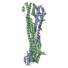
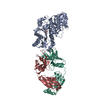
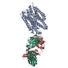
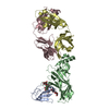
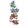
| Structure viewer | Molecule: MolmilJmol/JSmol |
|---|
- Downloads & links
-Download
| PDBx/mmCIF format | 4wic.cif.gz | 277.2 KB | Display | PDBx/mmCIF format |
|---|---|---|---|---|
| PDB format | pdb4wic.ent.gz | 228 KB | Display | PDB format |
| PDBx/mmJSON format | 4wic.json.gz | Tree view | PDBx/mmJSON format | |
| Others |  Other downloads Other downloads |
-Validation report
| Arichive directory | https://data.pdbj.org/pub/pdb/validation_reports/wi/4wicftp://data.pdbj.org/pub/pdb/validation_reports/wi/4wic | HTTPS FTP |
|---|
-Related structure data
| Similar structure data |
|---|


-Links
 PDBj
PDBj- Assembly
| Deposited unit | 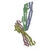
| ||||||||
|---|---|---|---|---|---|---|---|---|---|
| 1 | 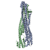
| ||||||||
| 2 | 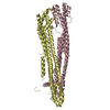
| ||||||||
| Unit cell |
|
-Components
| #1: Protein | Mass: 42145.477 Da / Num. of mol.: 4 Source method: isolated from a genetically manipulated source Source: (gene. exp.) Macacine herpesvirus 3 (Rhesus cytomegalovirus)Plasmid: pGEX-6P-1 / Production host:  Escherichia coli (E. coli) / Strain (production host): BL21(DE3) / References: UniProt: Q2FAE9 Escherichia coli (E. coli) / Strain (production host): BL21(DE3) / References: UniProt: Q2FAE9 |
|---|
-Experimental details
-Experiment
| Experiment | Method: X-RAY DIFFRACTION |
|---|
- Sample preparation
| Crystal | Density Matthews: 2.96 Å3/Da / Density % sol: 58.42 % |
|---|---|
| Crystal grow | Temperature: 293.15 K / Method: vapor diffusion, hanging drop Details: Reservoir 700 microL: 15 % (w/v) PEG 3350, 400 mM magnesium formate Drop ratio: 1 to 2 microL protein solution (20 mg/mL) plus 1 microL of reservoir solution supplemented with microseeds |
-Data collection
| Diffraction | Mean temperature: 100 K | ||||||||||||||||||
|---|---|---|---|---|---|---|---|---|---|---|---|---|---|---|---|---|---|---|---|
| Diffraction source | Source: SYNCHROTRON / Site: BESSY / Beamline: 14.1 / Wavelength: 0.9184, 1.0402, 1.0372, 1.0346, 0.9797 | ||||||||||||||||||
| Detector | Type: MARMOSAIC 225 mm CCD / Detector: CCD / Date: Sep 16, 2011 | ||||||||||||||||||
| Radiation | Protocol: MAD / Monochromatic (M) / Laue (L): M / Scattering type: x-ray | ||||||||||||||||||
| Radiation wavelength |
| ||||||||||||||||||
| Reflection | Resolution: 2.85→20 Å / Num. all: 45141 / Num. obs: 45137 / % possible obs: 99.5 % / Redundancy: 3.7 % / Biso Wilson estimate: 70.08 Å2 / Net I/σ(I): 16.9 | ||||||||||||||||||
| Reflection shell | Resolution: 2.85→2.92 Å / Redundancy: 1.7 % / % possible all: 99.7 |
- Processing
| Software |
| |||||||||||||||||||||||||||||||||||||||||||||||||||||||||||||||||||||||||||||||||||||||||||||||||||||||||||||||||||||||
|---|---|---|---|---|---|---|---|---|---|---|---|---|---|---|---|---|---|---|---|---|---|---|---|---|---|---|---|---|---|---|---|---|---|---|---|---|---|---|---|---|---|---|---|---|---|---|---|---|---|---|---|---|---|---|---|---|---|---|---|---|---|---|---|---|---|---|---|---|---|---|---|---|---|---|---|---|---|---|---|---|---|---|---|---|---|---|---|---|---|---|---|---|---|---|---|---|---|---|---|---|---|---|---|---|---|---|---|---|---|---|---|---|---|---|---|---|---|---|---|---|
| Refinement | Method to determine structure: MAD / Resolution: 2.85→19.935 Å / SU ML: 0.45 / Cross valid method: FREE R-VALUE / σ(F): 1.99 / Phase error: 31.23 / Stereochemistry target values: ML
| |||||||||||||||||||||||||||||||||||||||||||||||||||||||||||||||||||||||||||||||||||||||||||||||||||||||||||||||||||||||
| Solvent computation | Shrinkage radii: 0.9 Å / VDW probe radii: 1.11 Å / Solvent model: FLAT BULK SOLVENT MODEL | |||||||||||||||||||||||||||||||||||||||||||||||||||||||||||||||||||||||||||||||||||||||||||||||||||||||||||||||||||||||
| Displacement parameters | Biso mean: 75.6 Å2 | |||||||||||||||||||||||||||||||||||||||||||||||||||||||||||||||||||||||||||||||||||||||||||||||||||||||||||||||||||||||
| Refinement step | Cycle: LAST / Resolution: 2.85→19.935 Å
| |||||||||||||||||||||||||||||||||||||||||||||||||||||||||||||||||||||||||||||||||||||||||||||||||||||||||||||||||||||||
| Refine LS restraints |
| |||||||||||||||||||||||||||||||||||||||||||||||||||||||||||||||||||||||||||||||||||||||||||||||||||||||||||||||||||||||
| LS refinement shell |
|
