 Movie
Movie Controller

[English] 日本語
 Yorodumi
- PDB-4wfi: Crystal structure of PET-degrading cutinase Cut190 S226P mutant i... -
+ Open data

- Basic information
| Entry | Database: PDB / ID: 4wfi | ||||||
|---|---|---|---|---|---|---|---|
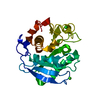
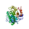
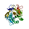
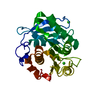
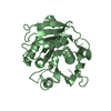
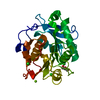
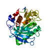
| Title | Crystal structure of PET-degrading cutinase Cut190 S226P mutant in Ca(2+)-free state | ||||||
 Components Components | Cutinase | ||||||
 Keywords Keywords | HYDROLASE / Cutinase / Polyesterase / alpha/beta-hydrolase fold | ||||||
| Function / homology |  Function and homology information Function and homology information | ||||||
| Biological species |  Saccharomonospora viridis (bacteria) Saccharomonospora viridis (bacteria) | ||||||
| Method | X-RAY DIFFRACTION / SYNCHROTRON / Resolution: 1.446 Å | ||||||
 Authors Authors | Miyakawa, T. / Mizushima, H. / Ohtsuka, J. / Oda, M. / Kawai, F. / Tanokura, M. | ||||||
 Citation Citation | Journal: Appl.Microbiol.Biotechnol. / Year: 2015 Title: Structural basis for the Ca(2+)-enhanced thermostability and activity of PET-degrading cutinase-like enzyme from Saccharomonospora viridis AHK190. Authors: Miyakawa, T. / Mizushima, H. / Ohtsuka, J. / Oda, M. / Kawai, F. / Tanokura, M. | ||||||
| History |
|
- Structure visualization
| Structure viewer | Molecule: MolmilJmol/JSmol |
|---|
- Downloads & links
-Download
| PDBx/mmCIF format | 4wfi.cif.gz | 64.7 KB | Display | PDBx/mmCIF format |
|---|---|---|---|---|
| PDB format | pdb4wfi.ent.gz | 49 KB | Display | PDB format |
| PDBx/mmJSON format | 4wfi.json.gz | Tree view | PDBx/mmJSON format | |
| Others |  Other downloads Other downloads |
-Validation report
| Arichive directory | https://data.pdbj.org/pub/pdb/validation_reports/wf/4wfiftp://data.pdbj.org/pub/pdb/validation_reports/wf/4wfi | HTTPS FTP |
|---|
-Related structure data


-Links
 PDBj
PDBj
- Assembly
| Deposited unit | 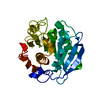
| ||||||||
|---|---|---|---|---|---|---|---|---|---|
| 1 |
| ||||||||
| Unit cell |
|
-Components
| #1: Protein | Mass: 30287.916 Da / Num. of mol.: 1 / Fragment: UNP RESIDUES 47-304 / Mutation: S226P Source method: isolated from a genetically manipulated source Source: (gene. exp.) Saccharomonospora viridis (bacteria) / Strain: AHK190 / Gene: Cut190 / Production host: Escherichia coli (E. coli) / Strain (production host): Rosetta-gami B (DE3) / References: UniProt: W0TJ64 |
|---|---|
| #2: Water | ChemComp-HOH / Water Mass: 18.015 Da / Num. of mol.: 226 / Source method: isolated from a natural source / Formula: H2O Mass: 18.015 Da / Num. of mol.: 226 / Source method: isolated from a natural source / Formula: H2O |
-Experimental details
-Experiment
| Experiment | Method: X-RAY DIFFRACTION |
|---|
- Sample preparation
| Crystal | Density Matthews: 2.15 Å3/Da / Density % sol: 42.77 % |
|---|---|
| Crystal grow | Temperature: 293 K / Method: vapor diffusion, sitting drop / pH: 7.5 Details: 50 mM Bis-Tris, 50 mM ammonium sulfate, 25% (w/v) pentaerythritol ethoxylate (15/4 EO/OH) |
-Data collection
| Diffraction | Mean temperature: 95 K |
|---|---|
| Diffraction source | Source: SYNCHROTRON / Site: Photon Factory  / Beamline: BL-17A / Wavelength: 1 Å / Beamline: BL-17A / Wavelength: 1 Å |
| Detector | Type: ADSC QUANTUM 270 / Detector: CCD / Date: Dec 2, 2012 |
| Radiation | Protocol: SINGLE WAVELENGTH / Monochromatic (M) / Laue (L): M / Scattering type: x-ray |
| Radiation wavelength | Wavelength: 1 Å / Relative weight: 1 |
| Reflection | Resolution: 1.446→20 Å / Num. obs: 47285 / % possible obs: 100 % / Redundancy: 7.2 % / Net I/σ(I): 74.7 |
- Processing
| Software | Name: PHENIX / Version: (phenix.refine: 1.9_1692) / Classification: refinement | ||||||||||||||||||||||||||||||||||||||||||||||||||||||||||||||||||||||||||||||||||||||||||||||||||||||||||||||||||||||||||||||
|---|---|---|---|---|---|---|---|---|---|---|---|---|---|---|---|---|---|---|---|---|---|---|---|---|---|---|---|---|---|---|---|---|---|---|---|---|---|---|---|---|---|---|---|---|---|---|---|---|---|---|---|---|---|---|---|---|---|---|---|---|---|---|---|---|---|---|---|---|---|---|---|---|---|---|---|---|---|---|---|---|---|---|---|---|---|---|---|---|---|---|---|---|---|---|---|---|---|---|---|---|---|---|---|---|---|---|---|---|---|---|---|---|---|---|---|---|---|---|---|---|---|---|---|---|---|---|---|
| Refinement | Resolution: 1.446→19.498 Å / SU ML: 0.13 / Cross valid method: FREE R-VALUE / σ(F): 0 / Phase error: 18.58 / Stereochemistry target values: ML
| ||||||||||||||||||||||||||||||||||||||||||||||||||||||||||||||||||||||||||||||||||||||||||||||||||||||||||||||||||||||||||||||
| Solvent computation | Shrinkage radii: 0.9 Å / VDW probe radii: 1.11 Å / Solvent model: FLAT BULK SOLVENT MODEL | ||||||||||||||||||||||||||||||||||||||||||||||||||||||||||||||||||||||||||||||||||||||||||||||||||||||||||||||||||||||||||||||
| Refinement step | Cycle: LAST / Resolution: 1.446→19.498 Å
| ||||||||||||||||||||||||||||||||||||||||||||||||||||||||||||||||||||||||||||||||||||||||||||||||||||||||||||||||||||||||||||||
| Refine LS restraints |
| ||||||||||||||||||||||||||||||||||||||||||||||||||||||||||||||||||||||||||||||||||||||||||||||||||||||||||||||||||||||||||||||
| LS refinement shell |
|
