| Entry | Database: PDB / ID: 4wei
|
|---|
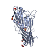
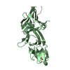
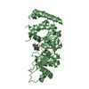
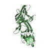
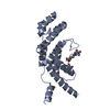
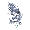
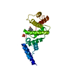
| Title | Crystal structure of the F4 fimbrial adhesin FaeG in complex with lactose |
|---|
 Components Components | K88 fimbrial protein AD |
|---|
 Keywords Keywords |  STRUCTURAL PROTEIN / Co-complex / Lectin / Adhesin / Immunoglobulin-like fold STRUCTURAL PROTEIN / Co-complex / Lectin / Adhesin / Immunoglobulin-like fold |
|---|
| Function / homology | pilus / alpha-lactose / K88 fimbrial protein AD Function and homology information Function and homology information |
|---|
| Biological species |   Escherichia coli (E. coli) Escherichia coli (E. coli) |
|---|
| Method | X-RAY DIFFRACTION / SYNCHROTRON / MOLECULAR REPLACEMENT / Resolution: 2.3 Å |
|---|
 Authors Authors | Moonens, K. / Van den Broeck, I. / De Kerpel, M. / Deboeck, F. / Raymaekers, H. / Remaut, H. / De Greve, H. |
|---|
| Funding support |  Belgium, 2items Belgium, 2items | Organization | Grant number | Country |
|---|
| FWO | G030411N | Belgium | | FWO - Hercules | UABR/09/005 | Belgium |
|
|---|
 Citation Citation | Journal: J.Biol.Chem. / Year: 2015
Title: Structural and Functional Insight into the Carbohydrate Receptor Binding of F4 Fimbriae-producing Enterotoxigenic Escherichia coli.
Authors: Moonens, K. / Van den Broeck, I. / De Kerpel, M. / Deboeck, F. / Raymaekers, H. / Remaut, H. / De Greve, H. |
|---|
| History | | Deposition | Sep 10, 2014 | Deposition site: RCSB / Processing site: PDBE |
|---|
| Revision 1.0 | Feb 4, 2015 | Provider: repository / Type: Initial release |
|---|
| Revision 1.1 | Feb 11, 2015 | Group: Database references |
|---|
| Revision 1.2 | Apr 8, 2015 | Group: Database references |
|---|
| Revision 2.0 | Jul 29, 2020 | Group: Atomic model / Data collection ...Atomic model / Data collection / Derived calculations / Structure summary
Category: atom_site / chem_comp ...atom_site / chem_comp / entity / entity_name_com / pdbx_branch_scheme / pdbx_chem_comp_identifier / pdbx_entity_branch / pdbx_entity_branch_descriptor / pdbx_entity_branch_link / pdbx_entity_branch_list / pdbx_entity_nonpoly / pdbx_molecule_features / pdbx_nonpoly_scheme / pdbx_struct_assembly_gen / pdbx_struct_mod_residue / pdbx_validate_chiral / struct_asym / struct_conn / struct_site / struct_site_gen
Item: _atom_site.B_iso_or_equiv / _atom_site.Cartn_x ..._atom_site.B_iso_or_equiv / _atom_site.Cartn_x / _atom_site.Cartn_y / _atom_site.Cartn_z / _atom_site.auth_asym_id / _atom_site.auth_atom_id / _atom_site.auth_comp_id / _atom_site.auth_seq_id / _atom_site.label_asym_id / _atom_site.label_atom_id / _atom_site.label_comp_id / _atom_site.label_entity_id / _atom_site.type_symbol / _chem_comp.name / _chem_comp.type / _pdbx_struct_assembly_gen.asym_id_list / _pdbx_struct_mod_residue.auth_asym_id / _pdbx_struct_mod_residue.auth_seq_id / _pdbx_struct_mod_residue.label_asym_id / _pdbx_validate_chiral.auth_asym_id / _pdbx_validate_chiral.auth_seq_id / _struct_conn.pdbx_dist_value / _struct_conn.pdbx_leaving_atom_flag / _struct_conn.pdbx_value_order / _struct_conn.ptnr1_auth_asym_id / _struct_conn.ptnr1_auth_seq_id / _struct_conn.ptnr1_label_atom_id / _struct_conn.ptnr2_auth_asym_id / _struct_conn.ptnr2_auth_seq_id / _struct_conn.ptnr2_label_asym_id / _struct_conn.ptnr2_label_atom_id
Description: Carbohydrate remediation / Provider: repository / Type: Remediation |
|---|
| Revision 2.1 | Jan 10, 2024 | Group: Data collection / Database references ...Data collection / Database references / Refinement description / Structure summary
Category: chem_comp / chem_comp_atom ...chem_comp / chem_comp_atom / chem_comp_bond / database_2 / pdbx_initial_refinement_model
Item: _chem_comp.pdbx_synonyms / _database_2.pdbx_DOI / _database_2.pdbx_database_accession |
|---|
|
|---|
 Movie
Movie Controller

 Yorodumi
 Open data
 Basic information
 Structure visualization
 Downloads & links
 Other downloads
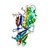


 PDBj
PDBj
 Assembly
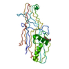


 Mass: 18.015 Da / Num. of mol.: 108 / Source method: isolated from a natural source / Formula: H2O
 Sample preparation
 / Beamline: PROXIMA 1 / Wavelength: 0.98 Å
 Processing