 Movie
Movie Controller

[English] 日本語
 Yorodumi
- PDB-4wcw: Ribosomal silencing factor during starvation or stationary phase ... -
+ Open data

- Basic information
| Entry | Database: PDB / ID: 4wcw | |||||||||
|---|---|---|---|---|---|---|---|---|---|---|
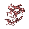
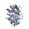
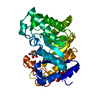
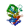
| Title | Ribosomal silencing factor during starvation or stationary phase (RsfS) from Mycobacterium tuberculosis | |||||||||
 Components Components | Ribosomal silencing factor RsfS | |||||||||
 Keywords Keywords |  TRANSLATION / Tuberculosis Ribosomal Silencing dimer / Structural Genomics / TB Structural Genomics Consortium / TBSGC TRANSLATION / Tuberculosis Ribosomal Silencing dimer / Structural Genomics / TB Structural Genomics Consortium / TBSGC | |||||||||
| Function / homology |  Function and homology information Function and homology informationnegative regulation of ribosome biogenesis / ribosomal large subunit binding / cytosolic ribosome assembly / negative regulation of translation / identical protein binding / cytoplasmSimilarity search - Function | |||||||||
| Biological species |   Mycobacterium tuberculosis (bacteria) Mycobacterium tuberculosis (bacteria) | |||||||||
| Method | X-RAY DIFFRACTION / SYNCHROTRON / MOLECULAR REPLACEMENT / Resolution: 2.1 Å | |||||||||
 Authors Authors | Li, X. / Sun, Q. / Jiang, C. / Yang, K. / Hung, L. / Zhang, J. / Sacchettini, J. / TB Structural Genomics Consortium (TBSGC) | |||||||||
| Funding support |  United States, 2items United States, 2items
| |||||||||
 Citation Citation | Journal: Structure / Year: 2015 Title: Structure of Ribosomal Silencing Factor Bound to Mycobacterium tuberculosis Ribosome. Authors: Xiaojun Li / Qingan Sun / Cai Jiang / Kailu Yang / Li-Wei Hung / Junjie Zhang / James C Sacchettini / Abstract: The ribosomal silencing factor RsfS slows cell growth by inhibiting protein synthesis during periods of diminished nutrient availability. The crystal structure of Mycobacterium tuberculosis (Mtb) ...The ribosomal silencing factor RsfS slows cell growth by inhibiting protein synthesis during periods of diminished nutrient availability. The crystal structure of Mycobacterium tuberculosis (Mtb) RsfS, together with the cryo-electron microscopy (EM) structure of the large subunit 50S of Mtb ribosome, reveals how inhibition of protein synthesis by RsfS occurs. RsfS binds to the 50S at L14, which, when occupied, blocks the association of the small subunit 30S. Although Mtb RsfS is a dimer in solution, only a single subunit binds to 50S. The overlap between the dimer interface and the L14 binding interface confirms that the RsfS dimer must first dissociate to a monomer in order to bind to L14. RsfS interacts primarily through electrostatic and hydrogen bonding to L14. The EM structure shows extended rRNA density that it is not found in the Escherichia coli ribosome, the most striking of these being the extended RNA helix of H54a. | |||||||||
| History |
|
- Structure visualization
| Structure viewer | Molecule: MolmilJmol/JSmol |
|---|
- Downloads & links
-Download
| PDBx/mmCIF format | 4wcw.cif.gz | 113.6 KB | Display | PDBx/mmCIF format |
|---|---|---|---|---|
| PDB format | pdb4wcw.ent.gz | 85.1 KB | Display | PDB format |
| PDBx/mmJSON format | 4wcw.json.gz | Tree view | PDBx/mmJSON format | |
| Others |  Other downloads Other downloads |
-Validation report
| Arichive directory | https://data.pdbj.org/pub/pdb/validation_reports/wc/4wcwftp://data.pdbj.org/pub/pdb/validation_reports/wc/4wcw | HTTPS FTP |
|---|
-Related structure data
| Related structure data | 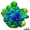 6177C  6178C  2o5aS S: Starting model for refinement C: citing same article ( |
|---|---|
| Similar structure data |


-Links
 PDBj
PDBj- Assembly
| Deposited unit | 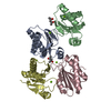
| ||||||||
|---|---|---|---|---|---|---|---|---|---|
| 1 | 
| ||||||||
| 2 | 
| ||||||||
| Unit cell |
| ||||||||
| Details | Dimer confirmed by gel filtration |
-Components
| #1: Protein | Mass: 15277.115 Da / Num. of mol.: 4 / Mutation: Y102A Source method: isolated from a genetically manipulated source Source: (gene. exp.) Mycobacterium tuberculosis (bacteria) / Strain: ATCC 25618 / H37Rv / Gene: rsfS, Rv2420c, P425_02518, RVBD_2420c / Production host: Mycobacterium smegmatis (bacteria) / References: UniProt: O86327#2: Chemical | ChemComp-MG /   Mass: 24.305 Da / Num. of mol.: 6 / Source method: obtained synthetically / Formula: Mg Mass: 24.305 Da / Num. of mol.: 6 / Source method: obtained synthetically / Formula: Mg#3: Chemical | 2-Methyl-2,4-pentanediol  Mass: 118.174 Da / Num. of mol.: 2 / Source method: obtained synthetically / Formula: C6H14O2 / Comment: precipitant*YM Mass: 118.174 Da / Num. of mol.: 2 / Source method: obtained synthetically / Formula: C6H14O2 / Comment: precipitant*YM#4: Water | ChemComp-HOH / | Water Mass: 18.015 Da / Num. of mol.: 378 / Source method: isolated from a natural source / Formula: H2O Mass: 18.015 Da / Num. of mol.: 378 / Source method: isolated from a natural source / Formula: H2O |
|---|
-Experimental details
-Experiment
| Experiment | Method: X-RAY DIFFRACTION |
|---|
- Sample preparation
| Crystal | Density Matthews: 2.33 Å3/Da / Density % sol: 47.13 % |
|---|---|
| Crystal grow | Temperature: 291 K / Method: vapor diffusion, sitting drop / pH: 6.5 Details: 0.1 M Sodium Cacodylate pH6.5, 0.2 M Magnesium Acetate, 30% MPD |
-Data collection
| Diffraction | Mean temperature: 120 K |
|---|---|
| Diffraction source | Source: SYNCHROTRON / Site: ALS / Beamline: 5.0.2 / Wavelength: 1 Å |
| Detector | Type: ADSC QUANTUM 315r / Detector: CCD / Date: Feb 4, 2014 |
| Radiation | Protocol: SINGLE WAVELENGTH / Monochromatic (M) / Laue (L): M / Scattering type: x-ray |
| Radiation wavelength | Wavelength: 1 Å / Relative weight: 1 |
| Reflection | Resolution: 2.1→46.06 Å / Num. obs: 49832 / % possible obs: 93.3 % / Redundancy: 1.7 % / Net I/σ(I): 6.3 |
- Processing
| Software | Name: PHENIX / Version: (phenix.refine: 1.8.2_1309) / Classification: refinement | |||||||||||||||||||||||||||||||||||||||||||||||||||||||||||||||||||||||||||||
|---|---|---|---|---|---|---|---|---|---|---|---|---|---|---|---|---|---|---|---|---|---|---|---|---|---|---|---|---|---|---|---|---|---|---|---|---|---|---|---|---|---|---|---|---|---|---|---|---|---|---|---|---|---|---|---|---|---|---|---|---|---|---|---|---|---|---|---|---|---|---|---|---|---|---|---|---|---|---|
| Refinement | Method to determine structure: MOLECULAR REPLACEMENT Starting model: 2O5A Resolution: 2.1→46.06 Å / SU ML: 0.24 / Cross valid method: FREE R-VALUE / σ(F): 2 / Phase error: 25.7 / Stereochemistry target values: ML
| |||||||||||||||||||||||||||||||||||||||||||||||||||||||||||||||||||||||||||||
| Solvent computation | Shrinkage radii: 0.9 Å / VDW probe radii: 1.11 Å / Solvent model: FLAT BULK SOLVENT MODEL | |||||||||||||||||||||||||||||||||||||||||||||||||||||||||||||||||||||||||||||
| Refinement step | Cycle: LAST / Resolution: 2.1→46.06 Å
| |||||||||||||||||||||||||||||||||||||||||||||||||||||||||||||||||||||||||||||
| Refine LS restraints |
| |||||||||||||||||||||||||||||||||||||||||||||||||||||||||||||||||||||||||||||
| LS refinement shell |
|
