 Movie
Movie Controller


+ Open data

- Basic information
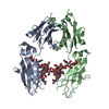
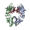
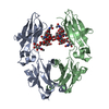
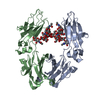
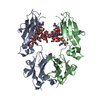
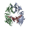
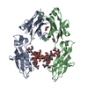
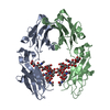
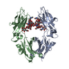
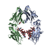
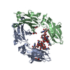
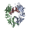
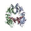
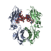
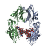
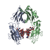
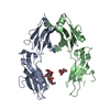
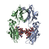
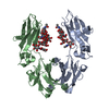
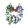
| Entry | Database: PDB / ID: 4w4n | |||||||||
|---|---|---|---|---|---|---|---|---|---|---|
| Title | Crystal structure of human Fc at 1.80 A | |||||||||
 Components Components | Ig gamma-1 chain C region | |||||||||
 Keywords Keywords |  IMMUNE SYSTEM / human Fc asymmetry dynamics immune molecule IMMUNE SYSTEM / human Fc asymmetry dynamics immune molecule | |||||||||
| Function / homology |  Function and homology informationcomplement-dependent cytotoxicity / antibody-dependent cellular cytotoxicity / Fc-gamma receptor I complex binding / Classical antibody-mediated complement activation / Initial triggering of complement / immunoglobulin complex, circulating / IgG immunoglobulin complex / immunoglobulin receptor binding / FCGR activation / Role of phospholipids in phagocytosis ...complement-dependent cytotoxicity / antibody-dependent cellular cytotoxicity / Fc-gamma receptor I complex binding / Classical antibody-mediated complement activation / Initial triggering of complement / immunoglobulin complex, circulating / IgG immunoglobulin complex / immunoglobulin receptor binding / FCGR activation / Role of phospholipids in phagocytosis / complement activation, classical pathway / antigen binding / FCGR3A-mediated IL10 synthesis / Regulation of Complement cascade / FCGR3A-mediated phagocytosis / B cell receptor signaling pathway / Regulation of actin dynamics for phagocytic cup formation / antibacterial humoral response / Interleukin-4 and Interleukin-13 signaling / blood microparticle / adaptive immune response / extracellular space / extracellular exosome / extracellular region / plasma membrane Function and homology informationcomplement-dependent cytotoxicity / antibody-dependent cellular cytotoxicity / Fc-gamma receptor I complex binding / Classical antibody-mediated complement activation / Initial triggering of complement / immunoglobulin complex, circulating / IgG immunoglobulin complex / immunoglobulin receptor binding / FCGR activation / Role of phospholipids in phagocytosis ...complement-dependent cytotoxicity / antibody-dependent cellular cytotoxicity / Fc-gamma receptor I complex binding / Classical antibody-mediated complement activation / Initial triggering of complement / immunoglobulin complex, circulating / IgG immunoglobulin complex / immunoglobulin receptor binding / FCGR activation / Role of phospholipids in phagocytosis / complement activation, classical pathway / antigen binding / FCGR3A-mediated IL10 synthesis / Regulation of Complement cascade / FCGR3A-mediated phagocytosis / B cell receptor signaling pathway / Regulation of actin dynamics for phagocytic cup formation / antibacterial humoral response / Interleukin-4 and Interleukin-13 signaling / blood microparticle / adaptive immune response / extracellular space / extracellular exosome / extracellular region / plasma membraneSimilarity search - Function | |||||||||
| Biological species |  Homo sapiens (human) Homo sapiens (human) | |||||||||
| Method | X-RAY DIFFRACTION / SYNCHROTRON / MOLECULAR REPLACEMENT / Resolution: 1.8 Å | |||||||||
 Authors Authors | Caaveiro, J.M.M. / Kiyoshi, M. / Tsumoto, K. | |||||||||
 Citation Citation | Journal: Nat Commun / Year: 2015 Title: Structural basis for binding of human IgG1 to its high-affinity human receptor Fc gamma RI Authors: Kiyoshi, M. / Caaveiro, J.M.M. / Kawai, T. / Tashiro, S. / Ide, T. / Asaoka, Y. / Hatayama, K. / Tsumoto, K. | |||||||||
| History |
|
- Structure visualization
| Structure viewer | Molecule: MolmilJmol/JSmol |
|---|
- Downloads & links
-Download
| PDBx/mmCIF format | 4w4n.cif.gz | 202.9 KB | Display | PDBx/mmCIF format |
|---|---|---|---|---|
| PDB format | pdb4w4n.ent.gz | 161.3 KB | Display | PDB format |
| PDBx/mmJSON format | 4w4n.json.gz | Tree view | PDBx/mmJSON format | |
| Others |  Other downloads Other downloads |
-Validation report
| Arichive directory | https://data.pdbj.org/pub/pdb/validation_reports/w4/4w4nftp://data.pdbj.org/pub/pdb/validation_reports/w4/4w4n | HTTPS FTP |
|---|
-Related structure data
| Related structure data | 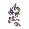 4w4oC  1l6xS C: citing same article ( S: Starting model for refinement |
|---|---|
| Similar structure data |
-Links
 PDBj
PDBj


- Assembly
| Deposited unit | 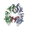
| ||||||||
|---|---|---|---|---|---|---|---|---|---|
| 1 |
| ||||||||
| Unit cell |
|
-Components
| #1: Protein | Mass: 25106.400 Da / Num. of mol.: 2 / Fragment: UNP residues 107-329 Source method: isolated from a genetically manipulated source Source: (gene. exp.) Homo sapiens (human)Description: THE PROTEIN IS COMMERCIALLY PURCHASED FROM ROCHE. Gene: IGHG1 / References: UniProt: P01857 #2: Polysaccharide | 2-acetamido-2-deoxy-beta-D-glucopyranose-(1-2)-alpha-D-mannopyranose-(1-3)-[2-acetamido-2-deoxy- ...2-acetamido-2-deoxy-beta-D-glucopyranose-(1-2)-alpha-D-mannopyranose-(1-3)-[2-acetamido-2-deoxy-beta-D-glucopyranose-(1-2)-alpha-D-mannopyranose-(1-6)]beta-D-mannopyranose-(1-4)-2-acetamido-2-deoxy-beta-D-glucopyranose-(1-4)-2-acetamido-2-deoxy-beta-D-glucopyranose | / Mass: 1317.209 Da / Num. of mol.: 1Source method: isolated from a genetically manipulated source #3: Polysaccharide | 2-acetamido-2-deoxy-beta-D-glucopyranose-(1-2)-alpha-D-mannopyranose-(1-3)-[2-acetamido-2-deoxy- ...2-acetamido-2-deoxy-beta-D-glucopyranose-(1-2)-alpha-D-mannopyranose-(1-3)-[2-acetamido-2-deoxy-beta-D-glucopyranose-(1-2)-alpha-D-mannopyranose-(1-6)]beta-D-mannopyranose-(1-4)-2-acetamido-2-deoxy-beta-D-glucopyranose-(1-4)-[alpha-L-fucopyranose-(1-6)]2-acetamido-2-deoxy-beta-D-glucopyranose | / Mass: 1463.349 Da / Num. of mol.: 1Source method: isolated from a genetically manipulated source #4: Chemical | ChemComp-GOL / | Glycerol  Mass: 92.094 Da / Num. of mol.: 1 / Source method: obtained synthetically / Formula: C3H8O3 Mass: 92.094 Da / Num. of mol.: 1 / Source method: obtained synthetically / Formula: C3H8O3#5: Water | ChemComp-HOH / | Water Mass: 18.015 Da / Num. of mol.: 374 / Source method: isolated from a natural source / Formula: H2O Mass: 18.015 Da / Num. of mol.: 374 / Source method: isolated from a natural source / Formula: H2O |
|---|
-Experimental details
-Experiment
| Experiment | Method: X-RAY DIFFRACTION / Number of used crystals: 1 |
|---|
- Sample preparation
| Crystal | Density Matthews: 2.72 Å3/Da / Density % sol: 54.8 % |
|---|---|
| Crystal grow | Temperature: 293 K / Method: vapor diffusion, hanging drop / pH: 6.1 Details: 0.2M calcium acetate, 0.1M sodium cacodylate, 19% PEG 8000 |
-Data collection
| Diffraction | Mean temperature: 100 K |
|---|---|
| Diffraction source | Source: SYNCHROTRON / Site: Photon Factory  / Beamline: BL-5A / Wavelength: 1 Å / Beamline: BL-5A / Wavelength: 1 Å |
| Detector | Type: ADSC QUANTUM 315 / Detector: CCD / Date: Feb 14, 2010 |
| Radiation | Protocol: SINGLE WAVELENGTH / Monochromatic (M) / Laue (L): M / Scattering type: x-ray |
| Radiation wavelength | Wavelength: 1 Å / Relative weight: 1 |
| Reflection | Resolution: 1.8→22.2 Å / Num. obs: 51158 / % possible obs: 99.1 % / Redundancy: 6.6 % / Rmerge(I) obs: 0.062 / Net I/σ(I): 18.4 |
| Reflection shell | Resolution: 1.8→1.9 Å / Redundancy: 6.4 % / Rmerge(I) obs: 0.787 / Mean I/σ(I) obs: 2.4 / % possible all: 97.3 |
- Processing
| Software | Name: REFMAC / Version: 5.8.0073 / Classification: refinement | ||||||||||||||||||||||||||||||||||||||||||||||||||||||||||||||||||||||||||||||||||||||||||||||||||||||||||||||||||||||||||||||||||||||||||||||||||||||||||||||||||||||||||||||||||||||
|---|---|---|---|---|---|---|---|---|---|---|---|---|---|---|---|---|---|---|---|---|---|---|---|---|---|---|---|---|---|---|---|---|---|---|---|---|---|---|---|---|---|---|---|---|---|---|---|---|---|---|---|---|---|---|---|---|---|---|---|---|---|---|---|---|---|---|---|---|---|---|---|---|---|---|---|---|---|---|---|---|---|---|---|---|---|---|---|---|---|---|---|---|---|---|---|---|---|---|---|---|---|---|---|---|---|---|---|---|---|---|---|---|---|---|---|---|---|---|---|---|---|---|---|---|---|---|---|---|---|---|---|---|---|---|---|---|---|---|---|---|---|---|---|---|---|---|---|---|---|---|---|---|---|---|---|---|---|---|---|---|---|---|---|---|---|---|---|---|---|---|---|---|---|---|---|---|---|---|---|---|---|---|---|
| Refinement | Method to determine structure: MOLECULAR REPLACEMENT Starting model: 1L6X Resolution: 1.8→22.188 Å / Cor.coef. Fo:Fc: 0.959 / Cor.coef. Fo:Fc free: 0.94 / SU B: 6.512 / SU ML: 0.106 / Cross valid method: THROUGHOUT / ESU R: 0.126 / ESU R Free: 0.127 / Stereochemistry target values: MAXIMUM LIKELIHOOD / Details: HYDROGENS HAVE BEEN USED IF PRESENT IN THE INPUT
| ||||||||||||||||||||||||||||||||||||||||||||||||||||||||||||||||||||||||||||||||||||||||||||||||||||||||||||||||||||||||||||||||||||||||||||||||||||||||||||||||||||||||||||||||||||||
| Solvent computation | Ion probe radii: 0.8 Å / Shrinkage radii: 0.8 Å / VDW probe radii: 1.2 Å / Solvent model: MASK | ||||||||||||||||||||||||||||||||||||||||||||||||||||||||||||||||||||||||||||||||||||||||||||||||||||||||||||||||||||||||||||||||||||||||||||||||||||||||||||||||||||||||||||||||||||||
| Displacement parameters | Biso mean: 43.233 Å2
| ||||||||||||||||||||||||||||||||||||||||||||||||||||||||||||||||||||||||||||||||||||||||||||||||||||||||||||||||||||||||||||||||||||||||||||||||||||||||||||||||||||||||||||||||||||||
| Refinement step | Cycle: 1 / Resolution: 1.8→22.188 Å
| ||||||||||||||||||||||||||||||||||||||||||||||||||||||||||||||||||||||||||||||||||||||||||||||||||||||||||||||||||||||||||||||||||||||||||||||||||||||||||||||||||||||||||||||||||||||
| Refine LS restraints |
|