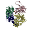
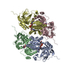
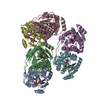
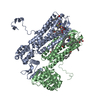
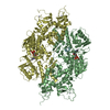
Entry Database : PDB / ID : 4v1uTitle Heterocyclase in complex with substrate and Cofactor Keywords / / Function / homology Function Domain/homology Component
/ / / / / / / / / Biological species LYNGBYA AESTUARII (bacteria)UNCULTURED PROCHLORON SP. (environmental samples)Method / / / Resolution : 2.86 Å Authors Koehnke, J. / Naismith, J.H. Journal : Nat.Chem.Biol. / Year : 2015Title : Structural Analysis of Leader Peptide Binding Enables Leader-Free Cyanobactin Processing.Authors : Koehnke, J. / Mann, G. / Bent, A.F. / Ludewig, H. / Shirran, S. / Botting, C. / Lebl, T. / Houssen, W.E. / Jaspars, M. / Naismith, J.H. History Deposition Oct 2, 2014 Deposition site / Processing site Revision 1.0 Jan 14, 2015 Provider / Type Revision 1.1 Jun 24, 2015 Group Revision 1.2 Jul 1, 2015 Group Revision 1.3 Aug 5, 2015 Group Revision 1.4 Jan 10, 2024 Group Data collection / Database references ... Data collection / Database references / Derived calculations / Other / Refinement description Category chem_comp_atom / chem_comp_bond ... chem_comp_atom / chem_comp_bond / database_2 / pdbx_database_status / pdbx_initial_refinement_model / pdbx_struct_conn_angle / struct_conn / struct_site Item _database_2.pdbx_DOI / _database_2.pdbx_database_accession ... _database_2.pdbx_DOI / _database_2.pdbx_database_accession / _pdbx_database_status.status_code_sf / _pdbx_struct_conn_angle.ptnr1_auth_seq_id / _pdbx_struct_conn_angle.ptnr1_label_atom_id / _pdbx_struct_conn_angle.ptnr1_label_seq_id / _pdbx_struct_conn_angle.ptnr3_auth_seq_id / _pdbx_struct_conn_angle.ptnr3_label_atom_id / _pdbx_struct_conn_angle.ptnr3_label_seq_id / _pdbx_struct_conn_angle.value / _struct_conn.pdbx_dist_value / _struct_conn.ptnr1_auth_comp_id / _struct_conn.ptnr1_auth_seq_id / _struct_conn.ptnr1_label_asym_id / _struct_conn.ptnr1_label_atom_id / _struct_conn.ptnr1_label_comp_id / _struct_conn.ptnr1_label_seq_id / _struct_conn.ptnr2_auth_comp_id / _struct_conn.ptnr2_auth_seq_id / _struct_conn.ptnr2_label_asym_id / _struct_conn.ptnr2_label_atom_id / _struct_conn.ptnr2_label_comp_id / _struct_conn.ptnr2_label_seq_id / _struct_site.pdbx_auth_asym_id / _struct_site.pdbx_auth_comp_id / _struct_site.pdbx_auth_seq_id
Show all Show less
 Movie
Movie Controller


 Open data
 Basic information
 Components
 Keywords
 HYDROLASE / HETEROCYCLASE / CYANOBACTINS
 Function and homology information
 LYNGBYA AESTUARII (bacteria)
 Authors
 Citation
 Structure visualization
 Downloads & links
 Other downloads


 PDBj
PDBj


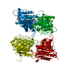
 Assembly
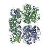


 Mass: 65.409 Da / Num. of mol.: 2 / Source method: obtained synthetically / Formula: Zn
 Mass: 347.221 Da / Num. of mol.: 2 / Source method: obtained synthetically / Formula: C10H14N5O7P / Comment: AMP*YM
 Mass: 40.078 Da / Num. of mol.: 4 / Source method: obtained synthetically / Formula: Ca
 Sample preparation
 / Beamline: I02 / Wavelength: 0.97949
  Processing