 Movie
Movie Controller

[English] 日本語
 Yorodumi
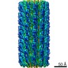
- PDB-4uzc: KSHV LANA (ORF73) C-terminal domain, spiral: hexagonal crystal form -
+ Open data

- Basic information
| Entry | Database: PDB / ID: 4uzc | ||||||
|---|---|---|---|---|---|---|---|
| Title | KSHV LANA (ORF73) C-terminal domain, spiral: hexagonal crystal form | ||||||
 Components Components | ORF 73 | ||||||
 Keywords Keywords |  VIRAL PROTEIN / DNA-BINDING DOMAIN / ORIGIN-BINDING DOMAIN / OLIGOMERIZATION DOMAIN / HHV-8 / GAMMAHERPESVIRUS / RHADINOVIRUS / PRIMARY EFFUSION LYMPHOMA / MULTICENTRIC CASTLEMAN'S DISEASE / TUMOR VIRUS / CANCER VIRAL PROTEIN / DNA-BINDING DOMAIN / ORIGIN-BINDING DOMAIN / OLIGOMERIZATION DOMAIN / HHV-8 / GAMMAHERPESVIRUS / RHADINOVIRUS / PRIMARY EFFUSION LYMPHOMA / MULTICENTRIC CASTLEMAN'S DISEASE / TUMOR VIRUS / CANCER | ||||||
| Function / homology |  Function and homology information Function and homology information | ||||||
| Biological species |   HUMAN HERPESVIRUS 8 HUMAN HERPESVIRUS 8 | ||||||
| Method | X-RAY DIFFRACTION / SYNCHROTRON / MOLECULAR REPLACEMENT / Resolution: 3.7 Å | ||||||
 Authors Authors | Hellert, J. / Krausze, J. / Luhrs, T. | ||||||
 Citation Citation | Journal: Proc.Natl.Acad.Sci.USA / Year: 2015 Title: The 3D Structure of Kaposi Sarcoma Herpesvirus Lana C-Terminal Domain Bound to DNA. Authors: Hellert, J. / Weidner-Glunde, M. / Krausze, J. / Lunsdorf, H. / Ritter, C. / Schulz, T.F. / Luhrs, T. | ||||||
| History |
| ||||||
| Remark 650 | HELIX DETERMINATION METHOD: AUTHOR PROVIDED. | ||||||
| Remark 700 | SHEET DETERMINATION METHOD: AUTHOR PROVIDED. |
- Structure visualization
| Structure viewer | Molecule: MolmilJmol/JSmol |
|---|
- Downloads & links
-Download
| PDBx/mmCIF format | 4uzc.cif.gz | 116.4 KB | Display | PDBx/mmCIF format |
|---|---|---|---|---|
| PDB format | pdb4uzc.ent.gz | 93.2 KB | Display | PDB format |
| PDBx/mmJSON format | 4uzc.json.gz | Tree view | PDBx/mmJSON format | |
| Others |  Other downloads Other downloads |
-Validation report
| Arichive directory | https://data.pdbj.org/pub/pdb/validation_reports/uz/4uzcftp://data.pdbj.org/pub/pdb/validation_reports/uz/4uzc | HTTPS FTP |
|---|
-Related structure data
| Related structure data |  4uzbC 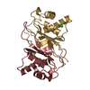 2ypyS C: citing same article ( S: Starting model for refinement |
|---|---|
| Similar structure data |
-Links
 PDBj
PDBj
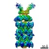
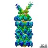
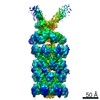
- Assembly
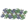
| Deposited unit | 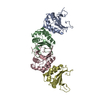
| ||||||||||||||||
|---|---|---|---|---|---|---|---|---|---|---|---|---|---|---|---|---|---|
| 1 | x 11 
| ||||||||||||||||
| Unit cell |
| ||||||||||||||||
| Noncrystallographic symmetry (NCS) | NCS oper:
| ||||||||||||||||
| Details | THE ASSEMBLY REPRESENTED IN THIS ENTRY HAS IRREGULAR HELICAL SYMMETRY WITH THE FOLLOWING AVERAGE PARAMETERS: ROTATION PER DIMER (TWIST) = -75.00 DEGREES RISE PER DIMER (HEIGHT) = 9.51 ANGSTROMS |
-Components
| #1: Protein | Mass: 15760.201 Da / Num. of mol.: 4 / Fragment: C-TERMINAL DOMAIN, RESIDUES 1013-1149 Source method: isolated from a genetically manipulated source Source: (gene. exp.) HUMAN HERPESVIRUS 8 / Plasmid: PET-BASED / Production host:  ESCHERICHIA COLI (E. coli) / Strain (production host): BL21(DE3) / References: UniProt: Q76SB0, UniProt: Q9QR71*PLUS ESCHERICHIA COLI (E. coli) / Strain (production host): BL21(DE3) / References: UniProt: Q76SB0, UniProt: Q9QR71*PLUS |
|---|
-Experimental details
-Experiment
| Experiment | Method: X-RAY DIFFRACTION / Number of used crystals: 1 |
|---|
- Sample preparation
| Crystal | Density Matthews: 2.79 Å3/Da / Density % sol: 55.88 % Description: DATA IN RESOLUTION RANGE 3.93 A - 3.87 A IS EXCLUDED DUE TO THE PRESENCE OF AN ICE RING |
|---|---|
| Crystal grow | Temperature: 293 K / Method: vapor diffusion, sitting drop / pH: 6.5 Details: 0.4 UL OF 31.5 MG/ML PROTEIN IN 5 MM BISTRIS-CL, 5 MM DTT, PH 6.5 WERE AS SUCH EQUILIBRATED AGAINST A RESERVOIR SOLUTION OF 170 MM LITHIUM ACETATE AND 18% (W/V) PEG3350 IN A SITTING DROP ...Details: 0.4 UL OF 31.5 MG/ML PROTEIN IN 5 MM BISTRIS-CL, 5 MM DTT, PH 6.5 WERE AS SUCH EQUILIBRATED AGAINST A RESERVOIR SOLUTION OF 170 MM LITHIUM ACETATE AND 18% (W/V) PEG3350 IN A SITTING DROP SETUP AT 20 DEGREE CENTIGRADE. AFTER TWO DAYS, CRYSTALS WERE DETACHED FROM THE CARRIER PLASTIC BY ADDING 1 UL OF 2 M AMMONIUM FORMATE. |
-Data collection
| Diffraction | Mean temperature: 100 K |
|---|---|
| Diffraction source | Source: SYNCHROTRON / Site: BESSY  / Beamline: 14.1 / Wavelength: 0.918409 / Beamline: 14.1 / Wavelength: 0.918409 |
| Detector | Type: DECTRIS PILATUS 6M / Detector: PIXEL / Date: Mar 19, 2014 / Details: MIRRORS |
| Radiation | Monochromator: SI(111) DOUBLE CRYSTAL / Protocol: SINGLE WAVELENGTH / Monochromatic (M) / Laue (L): M / Scattering type: x-ray |
| Radiation wavelength | Wavelength: 0.918409 Å / Relative weight: 1 |
| Reflection | Resolution: 3.7→89.36 Å / Num. obs: 7874 / % possible obs: 95.9 % / Observed criterion σ(I): 2 / Redundancy: 29.7 % / Biso Wilson estimate: 119.8 Å2 / Rmerge(I) obs: 0.23 / Net I/σ(I): 18.53 |
| Reflection shell | Resolution: 3.7→3.84 Å / Redundancy: 31.3 % / Mean I/σ(I) obs: 1.92 / % possible all: 99.1 |
- Processing
| Software |
| ||||||||||||||||||||||||||||||||||||||||||||||||||||||||||||||||||||||||||||||||||||||||||||||||||||||||||||||||||||||||||||||||||||||||||||||||||||||||||||||||||||||||||||||||||||||
|---|---|---|---|---|---|---|---|---|---|---|---|---|---|---|---|---|---|---|---|---|---|---|---|---|---|---|---|---|---|---|---|---|---|---|---|---|---|---|---|---|---|---|---|---|---|---|---|---|---|---|---|---|---|---|---|---|---|---|---|---|---|---|---|---|---|---|---|---|---|---|---|---|---|---|---|---|---|---|---|---|---|---|---|---|---|---|---|---|---|---|---|---|---|---|---|---|---|---|---|---|---|---|---|---|---|---|---|---|---|---|---|---|---|---|---|---|---|---|---|---|---|---|---|---|---|---|---|---|---|---|---|---|---|---|---|---|---|---|---|---|---|---|---|---|---|---|---|---|---|---|---|---|---|---|---|---|---|---|---|---|---|---|---|---|---|---|---|---|---|---|---|---|---|---|---|---|---|---|---|---|---|---|---|
| Refinement | Method to determine structure: MOLECULAR REPLACEMENT Starting model: PDB ENTRY 2YPY Resolution: 3.7→89.36 Å / Cor.coef. Fo:Fc: 0.936 / Cor.coef. Fo:Fc free: 0.908 / SU B: 40.346 / SU ML: 0.553 / Cross valid method: THROUGHOUT / ESU R Free: 0.692 / Stereochemistry target values: MAXIMUM LIKELIHOOD / Details: HYDROGENS HAVE BEEN ADDED IN THE RIDING POSITIONS.
| ||||||||||||||||||||||||||||||||||||||||||||||||||||||||||||||||||||||||||||||||||||||||||||||||||||||||||||||||||||||||||||||||||||||||||||||||||||||||||||||||||||||||||||||||||||||
| Solvent computation | Ion probe radii: 0.8 Å / Shrinkage radii: 0.8 Å / VDW probe radii: 1.2 Å / Solvent model: MASK | ||||||||||||||||||||||||||||||||||||||||||||||||||||||||||||||||||||||||||||||||||||||||||||||||||||||||||||||||||||||||||||||||||||||||||||||||||||||||||||||||||||||||||||||||||||||
| Displacement parameters | Biso mean: 115.815 Å2
| ||||||||||||||||||||||||||||||||||||||||||||||||||||||||||||||||||||||||||||||||||||||||||||||||||||||||||||||||||||||||||||||||||||||||||||||||||||||||||||||||||||||||||||||||||||||
| Refinement step | Cycle: LAST / Resolution: 3.7→89.36 Å
| ||||||||||||||||||||||||||||||||||||||||||||||||||||||||||||||||||||||||||||||||||||||||||||||||||||||||||||||||||||||||||||||||||||||||||||||||||||||||||||||||||||||||||||||||||||||
| Refine LS restraints |
|