 Movie
Movie Controller

[English] 日本語
 Yorodumi
- PDB-4uuy: Structural Identification of the Vps18 beta-propeller reveals a c... -
+ Open data

- Basic information
| Entry | Database: PDB / ID: 4uuy | ||||||
|---|---|---|---|---|---|---|---|
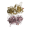
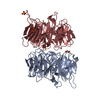
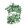
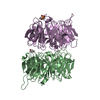
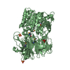
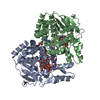
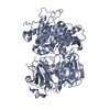
| Title | Structural Identification of the Vps18 beta-propeller reveals a critical role in the HOPS complex stability and function. | ||||||
 Components Components | VACUOLAR MEMBRANE PROTEIN PEP3 | ||||||
 Keywords Keywords |  TRANSPORT PROTEIN / HOPS / MEMBRANE FUSION / VACUOLE / ENDOSOME TRANSPORT PROTEIN / HOPS / MEMBRANE FUSION / VACUOLE / ENDOSOME | ||||||
| Function / homology |  Function and homology information Function and homology informationCORVET complex / organelle fusion / HOPS complex / regulation of SNARE complex assembly / vesicle tethering / regulation of vacuole fusion, non-autophagic / vacuole fusion, non-autophagic / Golgi to endosome transport / endosome organization / vacuole organization ...CORVET complex / organelle fusion / HOPS complex / regulation of SNARE complex assembly / vesicle tethering / regulation of vacuole fusion, non-autophagic / vacuole fusion, non-autophagic / Golgi to endosome transport / endosome organization / vacuole organization / vesicle docking involved in exocytosis / late endosome to vacuole transport / fungal-type vacuole membrane / intracellular protein transport / protein-macromolecule adaptor activity / early endosome membrane / endosome / metal ion binding / cytosolSimilarity search - Function | ||||||
| Biological species |  SACCHAROMYCES CEREVISIAE (brewer's yeast) SACCHAROMYCES CEREVISIAE (brewer's yeast) | ||||||
| Method | X-RAY DIFFRACTION / SYNCHROTRON / OTHER / Resolution: 2.14 Å | ||||||
 Authors Authors | Behrmann, H. / Gohlke, U. / Heinemann, U. | ||||||
 Citation Citation | Journal: J.Biol.Chem. / Year: 2014 Title: Structural Identification of the Vps18 Beta-Propeller Reveals a Critical Role in the Hops Complex Stability and Function. Authors: Behrmann, H. / Lurick, A. / Kuhlee, A. / Balderhaar, H.K. / Brocker, C. / Kummel, D. / Engelbrecht-Vandre, S. / Gohlke, U. / Raunser, S. / Heinemann, U. / Ungermann, C. | ||||||
| History |
|
- Structure visualization
| Structure viewer | Molecule: MolmilJmol/JSmol |
|---|
- Downloads & links
-Download
| PDBx/mmCIF format | 4uuy.cif.gz | 271 KB | Display | PDBx/mmCIF format |
|---|---|---|---|---|
| PDB format | pdb4uuy.ent.gz | 222.4 KB | Display | PDB format |
| PDBx/mmJSON format | 4uuy.json.gz | Tree view | PDBx/mmJSON format | |
| Others |  Other downloads Other downloads |
-Validation report
| Arichive directory | https://data.pdbj.org/pub/pdb/validation_reports/uu/4uuyftp://data.pdbj.org/pub/pdb/validation_reports/uu/4uuy | HTTPS FTP |
|---|
-Related structure data
| Similar structure data |
|---|


-Links
 PDBj
PDBj
- Assembly
| Deposited unit | 
| ||||||||
|---|---|---|---|---|---|---|---|---|---|
| 1 |
| ||||||||
| Unit cell |
| ||||||||
| Noncrystallographic symmetry (NCS) | NCS oper: (Code: given Matrix: (0.9491, 0.004047, 0.3151), Vector : |
-Components
| #1: Protein | Mass: 40405.297 Da / Num. of mol.: 2 / Fragment: BETA-PROPELLER, RESIDUES 2-348 Source method: isolated from a genetically manipulated source Source: (gene. exp.) SACCHAROMYCES CEREVISIAE (brewer's yeast)Plasmid: PQLINKH / Production host:  ESCHERICHIA COLI (E. coli) / Strain (production host): BL21(DE3) / Variant (production host): ROSETTA2 / References: UniProt: P27801 ESCHERICHIA COLI (E. coli) / Strain (production host): BL21(DE3) / Variant (production host): ROSETTA2 / References: UniProt: P27801#2: Chemical | ChemComp-SO4 / Sulfate  Mass: 96.063 Da / Num. of mol.: 6 / Source method: obtained synthetically / Formula: SO4 Mass: 96.063 Da / Num. of mol.: 6 / Source method: obtained synthetically / Formula: SO4#3: Chemical | Ethylene glycol  Mass: 62.068 Da / Num. of mol.: 3 / Source method: obtained synthetically / Formula: C2H6O2 Mass: 62.068 Da / Num. of mol.: 3 / Source method: obtained synthetically / Formula: C2H6O2#4: Chemical | Glycerol  Mass: 92.094 Da / Num. of mol.: 2 / Source method: obtained synthetically / Formula: C3H8O3 Mass: 92.094 Da / Num. of mol.: 2 / Source method: obtained synthetically / Formula: C3H8O3#5: Water | ChemComp-HOH / | Water Mass: 18.015 Da / Num. of mol.: 207 / Source method: isolated from a natural source / Formula: H2O Mass: 18.015 Da / Num. of mol.: 207 / Source method: isolated from a natural source / Formula: H2ONonpolymer details | ETHYLENE GLYCOL (EDO): SOLVENT GLYCEROL (GOL): SOLVENT SULPHATE (SO4): SOLVENT | Sequence details | FRAGMENT AA2-348 | |
|---|
-Experimental details
-Experiment
| Experiment | Method: X-RAY DIFFRACTION |
|---|
- Sample preparation
| Crystal | Density Matthews: 2.26 Å3/Da / Density % sol: 45.6 % / Description: NONE |
|---|---|
| Crystal grow | Details: 25 % (W/V) PEG 3350, 0.2 M (NH4)2SO4, 0.1 M BIS-TRIS, PH 6.5 |
-Data collection
| Diffraction | Mean temperature: 100 K |
|---|---|
| Diffraction source | Source: SYNCHROTRON / Site: BESSY  / Beamline: 14.1 / Wavelength: 0.91841 / Beamline: 14.1 / Wavelength: 0.91841 |
| Detector | Type: MARRESEARCH / Detector: CCD |
| Radiation | Protocol: SINGLE WAVELENGTH / Monochromatic (M) / Laue (L): M / Scattering type: x-ray |
| Radiation wavelength | Wavelength: 0.91841 Å / Relative weight: 1 |
| Reflection | Resolution: 2.14→63.12 Å / Num. obs: 39106 / % possible obs: 99.8 % / Observed criterion σ(I): 0 / Redundancy: 4.2 % / Biso Wilson estimate: 27 Å2 / Rmerge(I) obs: 0.11 / Net I/σ(I): 11.3 |
- Processing
| Software |
| ||||||||||||||||||||||||||||||||||||||||||||||||||||||||||||||||||||||||||||||||||||||||||||||||||||||||||||||||||||||||||||||||||||||||||||||||||||||||||||||||||||||||||||||||||||||
|---|---|---|---|---|---|---|---|---|---|---|---|---|---|---|---|---|---|---|---|---|---|---|---|---|---|---|---|---|---|---|---|---|---|---|---|---|---|---|---|---|---|---|---|---|---|---|---|---|---|---|---|---|---|---|---|---|---|---|---|---|---|---|---|---|---|---|---|---|---|---|---|---|---|---|---|---|---|---|---|---|---|---|---|---|---|---|---|---|---|---|---|---|---|---|---|---|---|---|---|---|---|---|---|---|---|---|---|---|---|---|---|---|---|---|---|---|---|---|---|---|---|---|---|---|---|---|---|---|---|---|---|---|---|---|---|---|---|---|---|---|---|---|---|---|---|---|---|---|---|---|---|---|---|---|---|---|---|---|---|---|---|---|---|---|---|---|---|---|---|---|---|---|---|---|---|---|---|---|---|---|---|---|---|
| Refinement | Method to determine structure: OTHER Starting model: NONE Resolution: 2.14→43.31 Å / Cor.coef. Fo:Fc: 0.943 / Cor.coef. Fo:Fc free: 0.913 / SU B: 12.945 / SU ML: 0.169 / Cross valid method: THROUGHOUT / ESU R: 0.276 / ESU R Free: 0.21 / Stereochemistry target values: MAXIMUM LIKELIHOOD Details: HYDROGENS HAVE BEEN ADDED IN THE RIDING POSITIONS. HYDROGENS HAVE BEEN ADDED IN THE RIDING POSITIONS WITH TLS ADDED
| ||||||||||||||||||||||||||||||||||||||||||||||||||||||||||||||||||||||||||||||||||||||||||||||||||||||||||||||||||||||||||||||||||||||||||||||||||||||||||||||||||||||||||||||||||||||
| Solvent computation | Ion probe radii: 0.8 Å / Shrinkage radii: 0.8 Å / VDW probe radii: 1.2 Å / Solvent model: MASK | ||||||||||||||||||||||||||||||||||||||||||||||||||||||||||||||||||||||||||||||||||||||||||||||||||||||||||||||||||||||||||||||||||||||||||||||||||||||||||||||||||||||||||||||||||||||
| Displacement parameters | Biso mean: 33.035 Å2
| ||||||||||||||||||||||||||||||||||||||||||||||||||||||||||||||||||||||||||||||||||||||||||||||||||||||||||||||||||||||||||||||||||||||||||||||||||||||||||||||||||||||||||||||||||||||
| Refinement step | Cycle: LAST / Resolution: 2.14→43.31 Å
| ||||||||||||||||||||||||||||||||||||||||||||||||||||||||||||||||||||||||||||||||||||||||||||||||||||||||||||||||||||||||||||||||||||||||||||||||||||||||||||||||||||||||||||||||||||||
| Refine LS restraints |
|