 Movie
Movie Controller

[English] 日本語
 Yorodumi
- PDB-4uu5: CRYSTAL STRUCTURE OF THE PDZ DOMAIN OF PALS1 IN COMPLEX WITH THE ... -
+ Open data

- Basic information
| Entry | Database: PDB / ID: 4uu5 | |||||||||
|---|---|---|---|---|---|---|---|---|---|---|
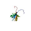
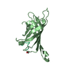
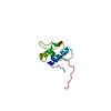
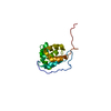
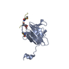
| Title | CRYSTAL STRUCTURE OF THE PDZ DOMAIN OF PALS1 IN COMPLEX WITH THE CRB PEPTIDE | |||||||||
 Components Components |
| |||||||||
 Keywords Keywords |  CELL ADHESION CELL ADHESION | |||||||||
| Function / homology |  Function and homology information Function and homology informationpost-embryonic retina morphogenesis in camera-type eye / protein localization to myelin sheath abaxonal region / SARS-CoV-1 targets PDZ proteins in cell-cell junction / establishment or maintenance of polarity of embryonic epithelium / myelin assembly / photoreceptor cell outer segment organization / morphogenesis of an epithelial sheet / Tight junction interactions / SARS-CoV-2 targets PDZ proteins in cell-cell junction / plasma membrane organization ...post-embryonic retina morphogenesis in camera-type eye / protein localization to myelin sheath abaxonal region / SARS-CoV-1 targets PDZ proteins in cell-cell junction / establishment or maintenance of polarity of embryonic epithelium / myelin assembly / photoreceptor cell outer segment organization / morphogenesis of an epithelial sheet / Tight junction interactions / SARS-CoV-2 targets PDZ proteins in cell-cell junction / plasma membrane organization / eye photoreceptor cell development / lateral loop / myelin sheath adaxonal region / regulation of transforming growth factor beta receptor signaling pathway / glial cell differentiation / Schmidt-Lanterman incisure / photoreceptor cell maintenance / peripheral nervous system myelin maintenance / establishment or maintenance of epithelial cell apical/basal polarity / heterophilic cell-cell adhesion via plasma membrane cell adhesion molecules / apical junction complex / generation of neurons / establishment or maintenance of cell polarity / central nervous system neuron development / microvillus / bicellular tight junction / blood vessel remodeling / visual perception / endoplasmic reticulum-Golgi intermediate compartment membrane / photoreceptor inner segment / protein localization to plasma membrane / adherens junction / cerebral cortex development / cell-cell signaling / gene expression / perikaryon / membrane => GO:0016020 / apical plasma membrane / protein domain specific binding / axon / calcium ion binding / Golgi apparatus / protein-containing complex / extracellular exosome / extracellular region / ATP binding / identical protein binding / plasma membrane / cytoplasmSimilarity search - Function | |||||||||
| Biological species |  HOMO SAPIENS (human) HOMO SAPIENS (human) | |||||||||
| Method | X-RAY DIFFRACTION / SYNCHROTRON / MOLECULAR REPLACEMENT / Resolution: 1.23 Å | |||||||||
 Authors Authors | Ivanova, M.E. / Purkiss, A.G. / McDonald, N.Q. | |||||||||
 Citation Citation | Journal: Acta Crystallogr. D Biol. Crystallogr. / Year: 2015 Title: Structures of the human Pals1 PDZ domain with and without ligand suggest gated access of Crb to the PDZ peptide-binding groove. Authors: Ivanova, M.E. / Fletcher, G.C. / O'Reilly, N. / Purkiss, A.G. / Thompson, B.J. / McDonald, N.Q. | |||||||||
| History |
|
- Structure visualization
| Structure viewer | Molecule: MolmilJmol/JSmol |
|---|
- Downloads & links
-Download
| PDBx/mmCIF format | 4uu5.cif.gz | 63.8 KB | Display | PDBx/mmCIF format |
|---|---|---|---|---|
| PDB format | pdb4uu5.ent.gz | 47 KB | Display | PDB format |
| PDBx/mmJSON format | 4uu5.json.gz | Tree view | PDBx/mmJSON format | |
| Others |  Other downloads Other downloads |
-Validation report
| Arichive directory | https://data.pdbj.org/pub/pdb/validation_reports/uu/4uu5ftp://data.pdbj.org/pub/pdb/validation_reports/uu/4uu5 | HTTPS FTP |
|---|
-Related structure data
| Related structure data |  4uu6C  1va8S C: citing same article ( S: Starting model for refinement |
|---|---|
| Similar structure data |


-Links
 PDBj
PDBj


- Assembly
| Deposited unit | 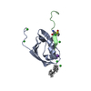
| ||||||||
|---|---|---|---|---|---|---|---|---|---|
| 1 |
| ||||||||
| Unit cell |
| ||||||||
| Components on special symmetry positions |
|
-Components
-Protein / Protein/peptide , 2 types, 2 molecules AB
| #1: Protein | Mass: 9650.056 Da / Num. of mol.: 1 / Fragment: PDZ DOMAIN, RESIDUES 251-335 Source method: isolated from a genetically manipulated source Source: (gene. exp.) HOMO SAPIENS (human) / Production host:  ESCHERICHIA COLI (E. coli) / Strain (production host): BL21 / References: UniProt: Q8N3R9 ESCHERICHIA COLI (E. coli) / Strain (production host): BL21 / References: UniProt: Q8N3R9 |
|---|---|
| #2: Protein/peptide | Mass: 2101.557 Da / Num. of mol.: 1 / Fragment: RESIDUES 1390-1406 Source method: isolated from a genetically manipulated source Source: (gene. exp.) HOMO SAPIENS (human) / Production host: ESCHERICHIA COLI (E. coli) / Strain (production host): BL21 / References: UniProt: P82279 |
-Non-polymers , 4 types, 130 molecules 


| #3: Chemical | ChemComp-CL / Chloride Mass: 35.453 Da / Num. of mol.: 8 / Source method: obtained synthetically / Formula: Cl Mass: 35.453 Da / Num. of mol.: 8 / Source method: obtained synthetically / Formula: Cl#4: Chemical | ChemComp-NA / |  Mass: 22.990 Da / Num. of mol.: 1 / Source method: obtained synthetically / Formula: Na Mass: 22.990 Da / Num. of mol.: 1 / Source method: obtained synthetically / Formula: Na#5: Chemical | ChemComp-PUN / | Isocetane Mass: 226.441 Da / Num. of mol.: 1 / Source method: obtained synthetically / Formula: C16H34 Mass: 226.441 Da / Num. of mol.: 1 / Source method: obtained synthetically / Formula: C16H34#6: Water | ChemComp-HOH / | WaterMass: 18.015 Da / Num. of mol.: 120 / Source method: isolated from a natural source / Formula: H2O |
|---|
-Details
| Sequence details | RESIDUES 251-335 N-TERMINAL RESIDUES GPLGS ARE VECTOR- DERIVED |
|---|
-Experimental details
-Experiment
| Experiment | Method: X-RAY DIFFRACTION / Number of used crystals: 1 |
|---|
- Sample preparation
| Crystal | Density Matthews: 2.64 Å3/Da / Density % sol: 53.37 % Description: MODEL WAS GENERATED BY PHYRE SERVER BASED ON 1VA8 |
|---|---|
| Crystal grow | pH: 7.5 / Details: 0.1M HEPES (PH 7.29), 2.68M NA CHLORIDE |
-Data collection
| Diffraction | Mean temperature: 100 K |
|---|---|
| Diffraction source | Source: SYNCHROTRON / Site: Diamond  / Beamline: I04-1 / Wavelength: 0.92 / Beamline: I04-1 / Wavelength: 0.92 |
| Detector | Type: DECTRIS PILATUS 6M / Detector: PIXEL / Date: Oct 25, 2013 / Details: MIRRORS |
| Radiation | Protocol: SINGLE WAVELENGTH / Monochromatic (M) / Laue (L): M / Scattering type: x-ray |
| Radiation wavelength | Wavelength: 0.92 Å / Relative weight: 1 |
| Reflection | Resolution: 1.23→52.84 Å / Num. obs: 35567 / % possible obs: 99.7 % / Observed criterion σ(I): 1.9 / Redundancy: 6.2 % / Biso Wilson estimate: 14.9 Å2 / Rmerge(I) obs: 0.04 / Net I/σ(I): 17.7 |
| Reflection shell | Resolution: 1.23→1.26 Å / Redundancy: 5.3 % / Rmerge(I) obs: 0.99 / Mean I/σ(I) obs: 1.9 / % possible all: 98.7 |
- Processing
| Software |
| ||||||||||||||||||||||||||||||||||||||||||||||||||||||||||||||||||||||||||||||||||||||||||||||||||
|---|---|---|---|---|---|---|---|---|---|---|---|---|---|---|---|---|---|---|---|---|---|---|---|---|---|---|---|---|---|---|---|---|---|---|---|---|---|---|---|---|---|---|---|---|---|---|---|---|---|---|---|---|---|---|---|---|---|---|---|---|---|---|---|---|---|---|---|---|---|---|---|---|---|---|---|---|---|---|---|---|---|---|---|---|---|---|---|---|---|---|---|---|---|---|---|---|---|---|---|
| Refinement | Method to determine structure: MOLECULAR REPLACEMENT Starting model: PDB ENTRY 1VA8 Resolution: 1.23→52.842 Å / SU ML: 0.11 / σ(F): 1.34 / Phase error: 16.77 / Stereochemistry target values: ML
| ||||||||||||||||||||||||||||||||||||||||||||||||||||||||||||||||||||||||||||||||||||||||||||||||||
| Solvent computation | Shrinkage radii: 0.9 Å / VDW probe radii: 1.11 Å / Solvent model: FLAT BULK SOLVENT MODEL | ||||||||||||||||||||||||||||||||||||||||||||||||||||||||||||||||||||||||||||||||||||||||||||||||||
| Refinement step | Cycle: LAST / Resolution: 1.23→52.842 Å
| ||||||||||||||||||||||||||||||||||||||||||||||||||||||||||||||||||||||||||||||||||||||||||||||||||
| Refine LS restraints |
| ||||||||||||||||||||||||||||||||||||||||||||||||||||||||||||||||||||||||||||||||||||||||||||||||||
| LS refinement shell |
|
