 Movie
Movie Controller


+ Open data

- Basic information
| Entry | Database: PDB / ID: 4urj | ||||||
|---|---|---|---|---|---|---|---|
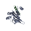
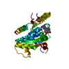
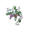
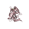
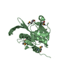
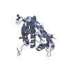
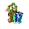
| Title | Crystal structure of human BJ-TSA-9 | ||||||
 Components Components | PROTEIN FAM83A | ||||||
 Keywords Keywords | UNKNOWN FUNCTION | ||||||
| Function / homology |  Function and homology information Function and homology informationphosphatidylinositol 3-kinase regulatory subunit binding / epidermal growth factor receptor signaling pathway / cell population proliferation /  protein kinase binding / signal transduction / identical protein binding / cytoplasm protein kinase binding / signal transduction / identical protein binding / cytoplasmSimilarity search - Function | ||||||
| Biological species |  HOMO SAPIENS (human) HOMO SAPIENS (human) | ||||||
| Method | X-RAY DIFFRACTION / SYNCHROTRON / MOLECULAR REPLACEMENT / Resolution: 2.68 Å | ||||||
 Authors Authors | Pinkas, D.M. / Sanvitale, C. / Wang, D. / Krojer, T. / Kopec, J. / Chaikuad, A. / Dixon Clarke, S. / Berridge, G. / Burgess-Brown, N. / von Delft, F. ...Pinkas, D.M. / Sanvitale, C. / Wang, D. / Krojer, T. / Kopec, J. / Chaikuad, A. / Dixon Clarke, S. / Berridge, G. / Burgess-Brown, N. / von Delft, F. / Arrowsmith, C. / Edwards, A. / Bountra, C. / Bullock, A. | ||||||
 Citation Citation | Journal: To be Published Title: Crystal Structure of Human Bj-Tsa-9 Authors: Pinkas, D.M. / Sanvitale, C. / Wang, D. / Krojer, T. / Kopec, J. / Chaikuad, A. / Dixon Clarke, S. / Berridge, G. / Burgess-Brown, N. / von Delft, F. / Arrowsmith, C. / Edwards, A. / Bountra, C. / Bullock, A. | ||||||
| History |
|
- Structure visualization
| Structure viewer | Molecule: MolmilJmol/JSmol |
|---|
- Downloads & links
-Download
| PDBx/mmCIF format | 4urj.cif.gz | 258.7 KB | Display | PDBx/mmCIF format |
|---|---|---|---|---|
| PDB format | pdb4urj.ent.gz | 213.1 KB | Display | PDB format |
| PDBx/mmJSON format | 4urj.json.gz | Tree view | PDBx/mmJSON format | |
| Others |  Other downloads Other downloads |
-Validation report
| Arichive directory | https://data.pdbj.org/pub/pdb/validation_reports/ur/4urjftp://data.pdbj.org/pub/pdb/validation_reports/ur/4urj | HTTPS FTP |
|---|
-Related structure data
| Similar structure data |
|---|


-Links
 PDBj
PDBj- Assembly
| Deposited unit | 
| ||||||||
|---|---|---|---|---|---|---|---|---|---|
| 1 | 
| ||||||||
| 2 | 
| ||||||||
| Unit cell |
|
-Components
| #1: Protein | Mass: 21168.578 Da / Num. of mol.: 4 / Fragment: DUF1669, UNP RESIDUES 122-304 Source method: isolated from a genetically manipulated source Source: (gene. exp.) HOMO SAPIENS (human) / Plasmid: PNIC28-BSA / Production host:  ESCHERICHIA COLI BL21(DE3) (bacteria) / Variant (production host): ROSETTA / References: UniProt: Q86UY5 ESCHERICHIA COLI BL21(DE3) (bacteria) / Variant (production host): ROSETTA / References: UniProt: Q86UY5#2: Chemical | ChemComp-EDO / Ethylene glycol  Mass: 62.068 Da / Num. of mol.: 7 / Source method: obtained synthetically / Formula: C2H6O2 Mass: 62.068 Da / Num. of mol.: 7 / Source method: obtained synthetically / Formula: C2H6O2#3: Water | ChemComp-HOH / | Water Mass: 18.015 Da / Num. of mol.: 103 / Source method: isolated from a natural source / Formula: H2O Mass: 18.015 Da / Num. of mol.: 103 / Source method: isolated from a natural source / Formula: H2O |
|---|
-Experimental details
-Experiment
| Experiment | Method: X-RAY DIFFRACTION / Number of used crystals: 1 |
|---|
- Sample preparation
| Crystal | Density Matthews: 2.7 Å3/Da / Density % sol: 54 % / Description: NONE |
|---|---|
| Crystal grow | Details: 0.8M SODIUM PHOSPHATE MONOBASIC, 0.8M SODIUM PHOSPHATE DIBASIC, 0.1M HEPES PH 7.5 |
-Data collection
| Diffraction | Mean temperature: 100 K |
|---|---|
| Diffraction source | Source: SYNCHROTRON / Site: Diamond  / Beamline: I03 / Wavelength: 0.97625 / Beamline: I03 / Wavelength: 0.97625 |
| Detector | Type: DECTRIS PILATUS 6M / Detector: PIXEL / Date: May 23, 2014 |
| Radiation | Protocol: SINGLE WAVELENGTH / Monochromatic (M) / Laue (L): M / Scattering type: x-ray |
| Radiation wavelength | Wavelength: 0.97625 Å / Relative weight: 1 |
| Reflection | Resolution: 2.68→42.08 Å / Num. obs: 25987 / % possible obs: 99.9 % / Observed criterion σ(I): -3 / Redundancy: 5.4 % / Biso Wilson estimate: 31.31 Å2 / Rmerge(I) obs: 0.2 / Net I/σ(I): 3.4 |
| Reflection shell | Resolution: 2.68→2.81 Å / Redundancy: 5.4 % / Rmerge(I) obs: 0.6 / Mean I/σ(I) obs: 1.9 / % possible all: 100 |
- Processing
| Software |
| |||||||||||||||||||||||||||||||||||||||||||||||||||||||||||||||||||||||||||||||||||||||||||||||||||||||||
|---|---|---|---|---|---|---|---|---|---|---|---|---|---|---|---|---|---|---|---|---|---|---|---|---|---|---|---|---|---|---|---|---|---|---|---|---|---|---|---|---|---|---|---|---|---|---|---|---|---|---|---|---|---|---|---|---|---|---|---|---|---|---|---|---|---|---|---|---|---|---|---|---|---|---|---|---|---|---|---|---|---|---|---|---|---|---|---|---|---|---|---|---|---|---|---|---|---|---|---|---|---|---|---|---|---|---|
| Refinement | Method to determine structure: MOLECULAR REPLACEMENT / Resolution: 2.68→42.084 Å / SU ML: 0.39 / σ(F): 1.97 / Phase error: 33.14 / Stereochemistry target values: ML
| |||||||||||||||||||||||||||||||||||||||||||||||||||||||||||||||||||||||||||||||||||||||||||||||||||||||||
| Solvent computation | Shrinkage radii: 0.9 Å / VDW probe radii: 1.11 Å / Solvent model: FLAT BULK SOLVENT MODEL | |||||||||||||||||||||||||||||||||||||||||||||||||||||||||||||||||||||||||||||||||||||||||||||||||||||||||
| Displacement parameters | Biso mean: 34.44 Å2 | |||||||||||||||||||||||||||||||||||||||||||||||||||||||||||||||||||||||||||||||||||||||||||||||||||||||||
| Refinement step | Cycle: LAST / Resolution: 2.68→42.084 Å
| |||||||||||||||||||||||||||||||||||||||||||||||||||||||||||||||||||||||||||||||||||||||||||||||||||||||||
| Refine LS restraints |
| |||||||||||||||||||||||||||||||||||||||||||||||||||||||||||||||||||||||||||||||||||||||||||||||||||||||||
| LS refinement shell |
|
