 Movie
Movie Controller


+ Open data

- Basic information
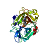
| Entry | Database: PDB / ID: 4unu | ||||||
|---|---|---|---|---|---|---|---|
| Title | MCG - a dimer of lambda variable domains | ||||||
 Components Components | IG LAMBDA CHAIN V-II REGION MGC | ||||||
 Keywords Keywords |  IMMUNE SYSTEM / BENCE-JONES / LIGHT CHAINS / IMMUNOGLOBULIN / AMYLOID IMMUNE SYSTEM / BENCE-JONES / LIGHT CHAINS / IMMUNOGLOBULIN / AMYLOID | ||||||
| Function / homology |  Function and homology information Function and homology informationCD22 mediated BCR regulation / Fc epsilon receptor (FCERI) signaling / immunoglobulin complex / Classical antibody-mediated complement activation / Initial triggering of complement / FCGR activation / Role of phospholipids in phagocytosis / Role of LAT2/NTAL/LAB on calcium mobilization / Scavenging of heme from plasma / FCERI mediated Ca+2 mobilization ...CD22 mediated BCR regulation / Fc epsilon receptor (FCERI) signaling / immunoglobulin complex / Classical antibody-mediated complement activation / Initial triggering of complement / FCGR activation / Role of phospholipids in phagocytosis / Role of LAT2/NTAL/LAB on calcium mobilization / Scavenging of heme from plasma / FCERI mediated Ca+2 mobilization / FCGR3A-mediated IL10 synthesis / Antigen activates B Cell Receptor (BCR) leading to generation of second messengers / antigen binding / Regulation of Complement cascade / Cell surface interactions at the vascular wall / FCERI mediated MAPK activation / FCGR3A-mediated phagocytosis / Regulation of actin dynamics for phagocytic cup formation / FCERI mediated NF-kB activation / Immunoregulatory interactions between a Lymphoid and a non-Lymphoid cell / adaptive immune response / Potential therapeutics for SARS / immune response / extracellular space / extracellular region / plasma membraneSimilarity search - Function | ||||||
| Biological species |  HOMO SAPIENS (human) HOMO SAPIENS (human) | ||||||
| Method | X-RAY DIFFRACTION / SYNCHROTRON / MOLECULAR REPLACEMENT / Resolution: 0.95 Å | ||||||
 Authors Authors | Brumshtein, B. / Esswein, S.R. / Landau, M. / Ryan, C. / Whitelegge, J. / Casio, D. / Sawaya, M.R. / Eisenberg, D.S. | ||||||
 Citation Citation | Journal: J.Biol.Chem. / Year: 2014 Title: Formation of Amyloid Fibers by Monomeric Light-Chain Variable Domains. Authors: Brumshtein, B. / Esswein, S.R. / Landau, M. / Ryan, C.M. / Whitelegge, J.P. / Phillips, M.L. / Cascio, D. / Sawaya, M.R. / Eisenberg, D.S. | ||||||
| History |
|
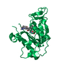
- Structure visualization
| Structure viewer | Molecule: MolmilJmol/JSmol |
|---|
- Downloads & links
-Download
| PDBx/mmCIF format | 4unu.cif.gz | 116.2 KB | Display | PDBx/mmCIF format |
|---|---|---|---|---|
| PDB format | pdb4unu.ent.gz | 88.4 KB | Display | PDB format |
| PDBx/mmJSON format | 4unu.json.gz | Tree view | PDBx/mmJSON format | |
| Others |  Other downloads Other downloads |
-Validation report
| Arichive directory | https://data.pdbj.org/pub/pdb/validation_reports/un/4unuftp://data.pdbj.org/pub/pdb/validation_reports/un/4unu | HTTPS FTP |
|---|
-Related structure data
| Related structure data |  4untC 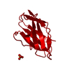 4unvC  3mcgS C: citing same article ( S: Starting model for refinement |
|---|---|
| Similar structure data |


-Links
 PDBj
PDBj


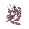
- Assembly
| Deposited unit | 
| ||||||||
|---|---|---|---|---|---|---|---|---|---|
| 1 |
| ||||||||
| Unit cell |
|
-Components
| #1: Antibody | Mass: 11565.510 Da / Num. of mol.: 2 / Fragment: LIGHT-CHAIN VARIABLE DOMAIN, RESIDUES 1-110 Source method: isolated from a genetically manipulated source Source: (gene. exp.) HOMO SAPIENS (human) / Tissue: BLOOD / Production host:  ESCHERICHIA COLI (E. coli) / Strain (production host): BL21(DE3) / References: UniProt: P01709 ESCHERICHIA COLI (E. coli) / Strain (production host): BL21(DE3) / References: UniProt: P01709#2: Chemical | ChemComp-15P / | Polyethylene glycol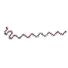  Mass: 1529.829 Da / Num. of mol.: 1 / Source method: obtained synthetically / Formula: C69H140O35 / Comment: precipitant*YM Mass: 1529.829 Da / Num. of mol.: 1 / Source method: obtained synthetically / Formula: C69H140O35 / Comment: precipitant*YM#3: Chemical | Sulfate  Mass: 96.063 Da / Num. of mol.: 3 / Source method: obtained synthetically / Formula: SO4 Mass: 96.063 Da / Num. of mol.: 3 / Source method: obtained synthetically / Formula: SO4#4: Water | ChemComp-HOH / | Water Mass: 18.015 Da / Num. of mol.: 373 / Source method: isolated from a natural source / Formula: H2O Mass: 18.015 Da / Num. of mol.: 373 / Source method: isolated from a natural source / Formula: H2O |
|---|
-Experimental details
-Experiment
| Experiment | Method: X-RAY DIFFRACTION / Number of used crystals: 1 |
|---|
- Sample preparation
| Crystal | Density Matthews: 1.8 Å3/Da / Density % sol: 31 % / Description: NONE |
|---|---|
| Crystal grow | pH: 8 / Details: 2M NACL, 2M (NH4)2SO4, pH 8 |
-Data collection
| Diffraction | Mean temperature: 100 K |
|---|---|
| Diffraction source | Source: SYNCHROTRON / Site: APS  / Beamline: 24-ID-C / Wavelength: 0.979 / Beamline: 24-ID-C / Wavelength: 0.979 |
| Detector | Type: DECTRIS PILATUS 6M-F / Detector: PIXEL / Date: Oct 26, 2012 |
| Radiation | Protocol: SINGLE WAVELENGTH / Monochromatic (M) / Laue (L): M / Scattering type: x-ray |
| Radiation wavelength | Wavelength: 0.979 Å / Relative weight: 1 |
| Reflection | Resolution: 0.95→40 Å / Num. obs: 89567 / % possible obs: 88 % / Observed criterion σ(I): 3 / Redundancy: 12 % / Rmerge(I) obs: 0.07 / Net I/σ(I): 21 |
| Reflection shell | Resolution: 0.95→0.97 Å / Redundancy: 3.2 % / Rmerge(I) obs: 0.7 / Mean I/σ(I) obs: 1.9 / % possible all: 34 |
- Processing
| Software |
| ||||||||||||||||||||||||||||||||||||||||||||||||||||||||||||||||||||||||||||||||||||||||||||||||||||||||||||||||||||||||||||||||||||||||||||||||||||||||||||||||||||||||||||||||||||||
|---|---|---|---|---|---|---|---|---|---|---|---|---|---|---|---|---|---|---|---|---|---|---|---|---|---|---|---|---|---|---|---|---|---|---|---|---|---|---|---|---|---|---|---|---|---|---|---|---|---|---|---|---|---|---|---|---|---|---|---|---|---|---|---|---|---|---|---|---|---|---|---|---|---|---|---|---|---|---|---|---|---|---|---|---|---|---|---|---|---|---|---|---|---|---|---|---|---|---|---|---|---|---|---|---|---|---|---|---|---|---|---|---|---|---|---|---|---|---|---|---|---|---|---|---|---|---|---|---|---|---|---|---|---|---|---|---|---|---|---|---|---|---|---|---|---|---|---|---|---|---|---|---|---|---|---|---|---|---|---|---|---|---|---|---|---|---|---|---|---|---|---|---|---|---|---|---|---|---|---|---|---|---|---|
| Refinement | Method to determine structure: MOLECULAR REPLACEMENT Starting model: PDB ENTRY 3MCG Resolution: 0.95→39.94 Å / Cor.coef. Fo:Fc: 0.984 / Cor.coef. Fo:Fc free: 0.977 / SU B: 0.454 / SU ML: 0.012 / Cross valid method: THROUGHOUT / ESU R: 0.019 / ESU R Free: 0.02 / Stereochemistry target values: MAXIMUM LIKELIHOOD / Details: HYDROGENS HAVE BEEN ADDED IN THE RIDING POSITIONS.
| ||||||||||||||||||||||||||||||||||||||||||||||||||||||||||||||||||||||||||||||||||||||||||||||||||||||||||||||||||||||||||||||||||||||||||||||||||||||||||||||||||||||||||||||||||||||
| Solvent computation | Ion probe radii: 0.8 Å / Shrinkage radii: 0.8 Å / VDW probe radii: 1.2 Å / Solvent model: MASK | ||||||||||||||||||||||||||||||||||||||||||||||||||||||||||||||||||||||||||||||||||||||||||||||||||||||||||||||||||||||||||||||||||||||||||||||||||||||||||||||||||||||||||||||||||||||
| Displacement parameters | Biso mean: 9.976 Å2
| ||||||||||||||||||||||||||||||||||||||||||||||||||||||||||||||||||||||||||||||||||||||||||||||||||||||||||||||||||||||||||||||||||||||||||||||||||||||||||||||||||||||||||||||||||||||
| Refinement step | Cycle: LAST / Resolution: 0.95→39.94 Å
| ||||||||||||||||||||||||||||||||||||||||||||||||||||||||||||||||||||||||||||||||||||||||||||||||||||||||||||||||||||||||||||||||||||||||||||||||||||||||||||||||||||||||||||||||||||||
| Refine LS restraints |
|