 Movie
Movie Controller

[English] 日本語
 Yorodumi
- PDB-4unk: Crystal structure of human triosephosphate isomerase (mutant N15D) -
+ Open data

- Basic information
| Entry | Database: PDB / ID: 4unk | ||||||
|---|---|---|---|---|---|---|---|
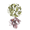
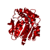
| Title | Crystal structure of human triosephosphate isomerase (mutant N15D) | ||||||
 Components Components | TRIOSEPHOSPHATE ISOMERASE | ||||||
 Keywords Keywords | ISOMERASE / DEAMIDATION | ||||||
| Function / homology |  Function and homology information Function and homology informationmethylglyoxal biosynthetic process / methylglyoxal synthase / methylglyoxal synthase activity / glyceraldehyde-3-phosphate biosynthetic process / glycerol catabolic process / triose-phosphate isomerase / triose-phosphate isomerase activity / Gluconeogenesis / canonical glycolysis / Glycolysis ...methylglyoxal biosynthetic process / methylglyoxal synthase / methylglyoxal synthase activity / glyceraldehyde-3-phosphate biosynthetic process / glycerol catabolic process / triose-phosphate isomerase / triose-phosphate isomerase activity / Gluconeogenesis / canonical glycolysis / Glycolysis / gluconeogenesis / glycolytic process / ubiquitin protein ligase binding / protein homodimerization activity / extracellular space / extracellular exosome / nucleus / cytosolSimilarity search - Function | ||||||
| Biological species |  HOMO SAPIENS (human) HOMO SAPIENS (human) | ||||||
| Method | X-RAY DIFFRACTION / SYNCHROTRON / MOLECULAR REPLACEMENT / Resolution: 2 Å | ||||||
 Authors Authors | DeLaMora-DeLaMora, I. / Torres-Larios, A. / Enriquez-Flores, S. / Mendez, S.T. / Castillo-Villanueva, A. / Gomez-Manzo, S. / Lopez-Velazquez, G. / Marcial-Quino, J. / Torres-Arroyo, A. / Garcia-Torres, I. ...DeLaMora-DeLaMora, I. / Torres-Larios, A. / Enriquez-Flores, S. / Mendez, S.T. / Castillo-Villanueva, A. / Gomez-Manzo, S. / Lopez-Velazquez, G. / Marcial-Quino, J. / Torres-Arroyo, A. / Garcia-Torres, I. / Reyes-Vivas, H. / Oria-Hernandez, J. | ||||||
 Citation Citation | Journal: To be Published Title: Crystal Structure of Human Triosephosphate Isomerase (Mutant N15D) Authors: Delamora-Delamora, I. / Torres-Larios, A. / Enriquez-Flores, S. / Mendez, S.T. / Castillo-Villanueva, A. / Gomez-Manzo, S. / Lopez-Velazquez, G. / Marcial-Quino, J. / Torres-Arroyo, A. / ...Authors: Delamora-Delamora, I. / Torres-Larios, A. / Enriquez-Flores, S. / Mendez, S.T. / Castillo-Villanueva, A. / Gomez-Manzo, S. / Lopez-Velazquez, G. / Marcial-Quino, J. / Torres-Arroyo, A. / Garcia-Torres, I. / Reyes-Vivas, H. / Oria-Hernandez, J. | ||||||
| History |
| ||||||
| Remark 700 | SHEET DETERMINATION METHOD: DSSP THE SHEETS PRESENTED AS "AA" IN EACH CHAIN ON SHEET RECORDS BELOW ... SHEET DETERMINATION METHOD: DSSP THE SHEETS PRESENTED AS "AA" IN EACH CHAIN ON SHEET RECORDS BELOW IS ACTUALLY AN 8-STRANDED BARREL THIS IS REPRESENTED BY A 9-STRANDED SHEET IN WHICH THE FIRST AND LAST STRANDS ARE IDENTICAL. THE SHEETS PRESENTED AS "BA" IN EACH CHAIN ON SHEET RECORDS BELOW IS ACTUALLY AN 8-STRANDED BARREL THIS IS REPRESENTED BY A 9-STRANDED SHEET IN WHICH THE FIRST AND LAST STRANDS ARE IDENTICAL. |
- Structure visualization
| Structure viewer | Molecule: MolmilJmol/JSmol |
|---|
- Downloads & links
-Download
| PDBx/mmCIF format | 4unk.cif.gz | 199.1 KB | Display | PDBx/mmCIF format |
|---|---|---|---|---|
| PDB format | pdb4unk.ent.gz | 162.4 KB | Display | PDB format |
| PDBx/mmJSON format | 4unk.json.gz | Tree view | PDBx/mmJSON format | |
| Others |  Other downloads Other downloads |
-Validation report
| Arichive directory | https://data.pdbj.org/pub/pdb/validation_reports/un/4unkftp://data.pdbj.org/pub/pdb/validation_reports/un/4unk | HTTPS FTP |
|---|
-Related structure data
| Related structure data |  2jk2S S: Starting model for refinement |
|---|---|
| Similar structure data |


-Links
 PDBj
PDBj

- Assembly
| Deposited unit | 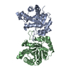
| ||||||||
|---|---|---|---|---|---|---|---|---|---|
| 1 |
| ||||||||
| Unit cell |
| ||||||||
| Noncrystallographic symmetry (NCS) | NCS oper: (Code: given Matrix: (-0.6361, 0.2254, -0.7379), Vector : |
-Components
| #1: Protein | / TIM / TRIOSE-PHOSPHATE ISOMERASE Mass: 26715.385 Da / Num. of mol.: 2 / Fragment: RESIDUES 39-286 / Mutation: YES Source method: isolated from a genetically manipulated source Source: (gene. exp.) HOMO SAPIENS (human) / Production host:  ESCHERICHIA COLI (E. coli) / Strain (production host): BL21(DE3) / Variant (production host): CODON PLUS RIL / References: UniProt: P60174, triose-phosphate isomerase ESCHERICHIA COLI (E. coli) / Strain (production host): BL21(DE3) / Variant (production host): CODON PLUS RIL / References: UniProt: P60174, triose-phosphate isomerase#2: Water | ChemComp-HOH / | Water Mass: 18.015 Da / Num. of mol.: 452 / Source method: isolated from a natural source / Formula: H2O Mass: 18.015 Da / Num. of mol.: 452 / Source method: isolated from a natural source / Formula: H2O |
|---|
-Experimental details
-Experiment
| Experiment | Method: X-RAY DIFFRACTION / Number of used crystals: 1 |
|---|
- Sample preparation
| Crystal | Density Matthews: 2.12 Å3/Da / Density % sol: 41.54 % / Description: NONE |
|---|---|
| Crystal grow | pH: 8.5 Details: 200 MM AMMONIUM ACETATE, 100 MM TRIS PH 8.5, 25% V/VW/V POLYETHYLENE GLYCOL 3350 |
-Data collection
| Diffraction | Mean temperature: 110 K |
|---|---|
| Diffraction source | Source: SYNCHROTRON / Site: APS  / Beamline: 21-ID-F / Wavelength: 0.97872 / Beamline: 21-ID-F / Wavelength: 0.97872 |
| Detector | Type: MARRESEARCH / Detector: CCD / Date: Jul 28, 2010 / Details: MIRRORS |
| Radiation | Monochromator: DIAMOND / Protocol: SINGLE WAVELENGTH / Monochromatic (M) / Laue (L): M / Scattering type: x-ray |
| Radiation wavelength | Wavelength: 0.97872 Å / Relative weight: 1 |
| Reflection | Resolution: 2→48.81 Å / Num. obs: 34112 / % possible obs: 99.7 % / Observed criterion σ(I): 0 / Redundancy: 3.5 % / Biso Wilson estimate: 31.06 Å2 / Rmerge(I) obs: 0.07 / Net I/σ(I): 10.8 |
| Reflection shell | Resolution: 2→2.11 Å / Redundancy: 3.5 % / Rmerge(I) obs: 0.29 / Mean I/σ(I) obs: 3.6 / % possible all: 100 |
- Processing
| Software |
| ||||||||||||||||||||||||||||||||||||||||||||||||||||||||||||||||||||||||||||||||||||||||||||||||||||||||||||||||||||||||||||||||||||||||||||||||||||||||||||||||||||||||||||||||||||||||||||||||||||||||
|---|---|---|---|---|---|---|---|---|---|---|---|---|---|---|---|---|---|---|---|---|---|---|---|---|---|---|---|---|---|---|---|---|---|---|---|---|---|---|---|---|---|---|---|---|---|---|---|---|---|---|---|---|---|---|---|---|---|---|---|---|---|---|---|---|---|---|---|---|---|---|---|---|---|---|---|---|---|---|---|---|---|---|---|---|---|---|---|---|---|---|---|---|---|---|---|---|---|---|---|---|---|---|---|---|---|---|---|---|---|---|---|---|---|---|---|---|---|---|---|---|---|---|---|---|---|---|---|---|---|---|---|---|---|---|---|---|---|---|---|---|---|---|---|---|---|---|---|---|---|---|---|---|---|---|---|---|---|---|---|---|---|---|---|---|---|---|---|---|---|---|---|---|---|---|---|---|---|---|---|---|---|---|---|---|---|---|---|---|---|---|---|---|---|---|---|---|---|---|---|---|---|
| Refinement | Method to determine structure: MOLECULAR REPLACEMENT Starting model: PDB ENTRY 2JK2 Resolution: 2→45.292 Å / SU ML: 0.23 / σ(F): 1.35 / Phase error: 21.21 / Stereochemistry target values: ML Details: RESIDUES 171-176 OF CHAIN B, KNOWN AS LOOP 6, ARE DISORDERED.
| ||||||||||||||||||||||||||||||||||||||||||||||||||||||||||||||||||||||||||||||||||||||||||||||||||||||||||||||||||||||||||||||||||||||||||||||||||||||||||||||||||||||||||||||||||||||||||||||||||||||||
| Solvent computation | Shrinkage radii: 0.9 Å / VDW probe radii: 1.11 Å / Solvent model: FLAT BULK SOLVENT MODEL | ||||||||||||||||||||||||||||||||||||||||||||||||||||||||||||||||||||||||||||||||||||||||||||||||||||||||||||||||||||||||||||||||||||||||||||||||||||||||||||||||||||||||||||||||||||||||||||||||||||||||
| Displacement parameters | Biso mean: 30.4 Å2 | ||||||||||||||||||||||||||||||||||||||||||||||||||||||||||||||||||||||||||||||||||||||||||||||||||||||||||||||||||||||||||||||||||||||||||||||||||||||||||||||||||||||||||||||||||||||||||||||||||||||||
| Refinement step | Cycle: LAST / Resolution: 2→45.292 Å
| ||||||||||||||||||||||||||||||||||||||||||||||||||||||||||||||||||||||||||||||||||||||||||||||||||||||||||||||||||||||||||||||||||||||||||||||||||||||||||||||||||||||||||||||||||||||||||||||||||||||||
| Refine LS restraints |
| ||||||||||||||||||||||||||||||||||||||||||||||||||||||||||||||||||||||||||||||||||||||||||||||||||||||||||||||||||||||||||||||||||||||||||||||||||||||||||||||||||||||||||||||||||||||||||||||||||||||||
| LS refinement shell |
| ||||||||||||||||||||||||||||||||||||||||||||||||||||||||||||||||||||||||||||||||||||||||||||||||||||||||||||||||||||||||||||||||||||||||||||||||||||||||||||||||||||||||||||||||||||||||||||||||||||||||
| Refinement TLS params. | Method: refined / Refine-ID: X-RAY DIFFRACTION
| ||||||||||||||||||||||||||||||||||||||||||||||||||||||||||||||||||||||||||||||||||||||||||||||||||||||||||||||||||||||||||||||||||||||||||||||||||||||||||||||||||||||||||||||||||||||||||||||||||||||||
| Refinement TLS group |
|
