 Movie
Movie Controller

[English] 日本語
 Yorodumi
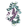
- PDB-4umn: Structure of a stapled peptide antagonist bound to Nutlin-resista... -
+ Open data

- Basic information
| Entry | Database: PDB / ID: 4umn | ||||||||||||
|---|---|---|---|---|---|---|---|---|---|---|---|---|---|
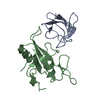
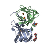
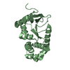
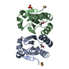
| Title | Structure of a stapled peptide antagonist bound to Nutlin-resistant Mdm2. | ||||||||||||
 Components Components |
| ||||||||||||
 Keywords Keywords |  CELL CYCLE CELL CYCLE | ||||||||||||
| Function / homology |  Function and homology information Function and homology informationcellular response to vitamin B1 / response to formaldehyde / response to water-immersion restraint stress / response to ether / traversing start control point of mitotic cell cycle / negative regulation of intrinsic apoptotic signaling pathway by p53 class mediator / negative regulation of signal transduction by p53 class mediator / fibroblast activation / atrial septum development / receptor serine/threonine kinase binding ...cellular response to vitamin B1 / response to formaldehyde / response to water-immersion restraint stress / response to ether / traversing start control point of mitotic cell cycle / negative regulation of intrinsic apoptotic signaling pathway by p53 class mediator / negative regulation of signal transduction by p53 class mediator / fibroblast activation / atrial septum development / receptor serine/threonine kinase binding / Trafficking of AMPA receptors / positive regulation of vascular associated smooth muscle cell migration / response to iron ion / peroxisome proliferator activated receptor binding / negative regulation of protein processing / response to steroid hormone / SUMO transferase activity / NEDD8 ligase activity / AKT phosphorylates targets in the cytosol / cellular response to peptide hormone stimulus / atrioventricular valve morphogenesis / ventricular septum development / endocardial cushion morphogenesis / positive regulation of muscle cell differentiation / SUMOylation of ubiquitinylation proteins / cellular response to alkaloid / blood vessel development / regulation of protein catabolic process / cardiac septum morphogenesis / Constitutive Signaling by AKT1 E17K in Cancer / negative regulation of DNA damage response, signal transduction by p53 class mediator / response to magnesium ion / ligase activity / protein sumoylation / SUMOylation of transcription factors / protein localization to nucleus / cellular response to actinomycin D / cellular response to UV-C / blood vessel remodeling / cellular response to estrogen stimulus / protein autoubiquitination / ribonucleoprotein complex binding / DNA damage response, signal transduction by p53 class mediator resulting in cell cycle arrest / positive regulation of vascular associated smooth muscle cell proliferation / transcription repressor complex / NPAS4 regulates expression of target genes / regulation of heart rate / proteolysis involved in protein catabolic process / positive regulation of mitotic cell cycle / positive regulation of protein export from nucleus / response to cocaine / ubiquitin binding / Stabilization of p53 / Regulation of RUNX3 expression and activity / protein destabilization / cellular response to gamma radiation / RING-type E3 ubiquitin transferase / establishment of protein localization / Oncogene Induced Senescence / Regulation of TP53 Activity through Methylation / response to toxic substance / cellular response to hydrogen peroxide / cellular response to growth factor stimulus / protein polyubiquitination / ubiquitin-protein transferase activity / endocytic vesicle membrane / ubiquitin protein ligase activity / disordered domain specific binding / Regulation of TP53 Degradation / positive regulation of proteasomal ubiquitin-dependent protein catabolic process / p53 binding / negative regulation of neuron projection development / 5S rRNA binding / ubiquitin-dependent protein catabolic process / cellular response to hypoxia / regulation of gene expression / proteasome-mediated ubiquitin-dependent protein catabolic process / protein-containing complex assembly / Oxidative Stress Induced Senescence / Regulation of TP53 Activity through Phosphorylation / amyloid fibril formation / protein ubiquitination / regulation of cell cycle / Ub-specific processing proteases / response to xenobiotic stimulus / protein domain specific binding / response to antibiotic / negative regulation of DNA-templated transcription / apoptotic process / ubiquitin protein ligase binding / positive regulation of cell population proliferation / positive regulation of gene expression / nucleolus / negative regulation of apoptotic process / negative regulation of transcription by RNA polymerase II / enzyme binding / protein-containing complex / zinc ion binding / nucleoplasm / identical protein bindingSimilarity search - Function | ||||||||||||
| Biological species |  Homo sapiens (human) Homo sapiens (human)SYNTHETIC CONSTRUCT (others) | ||||||||||||
| Method | X-RAY DIFFRACTION / SYNCHROTRON / MOLECULAR REPLACEMENT / Resolution: 1.99 Å | ||||||||||||
 Authors Authors | Chee, S. / Wongsantichon, J. / Quah, S. / Robinson, R.C. / Verma, C. / Lane, D.P. / Brown, C.J. / Ghadessy, F.J. | ||||||||||||
 Citation Citation | Journal: PLoS ONE / Year: 2014 Title: Structure of a stapled peptide antagonist bound to nutlin-resistant Mdm2. Authors: Chee, S.M. / Wongsantichon, J. / Soo Tng, Q. / Robinson, R. / Joseph, T.L. / Verma, C. / Lane, D.P. / Brown, C.J. / Ghadessy, F.J. | ||||||||||||
| History |
|
- Structure visualization
| Structure viewer | Molecule: MolmilJmol/JSmol |
|---|
- Downloads & links
-Download
| PDBx/mmCIF format | 4umn.cif.gz | 105 KB | Display | PDBx/mmCIF format |
|---|---|---|---|---|
| PDB format | pdb4umn.ent.gz | 81.1 KB | Display | PDB format |
| PDBx/mmJSON format | 4umn.json.gz | Tree view | PDBx/mmJSON format | |
| Others |  Other downloads Other downloads |
-Validation report
| Arichive directory | https://data.pdbj.org/pub/pdb/validation_reports/um/4umnftp://data.pdbj.org/pub/pdb/validation_reports/um/4umn | HTTPS FTP |
|---|
-Related structure data
| Related structure data |  2axiS S: Starting model for refinement |
|---|---|
| Similar structure data |


-Links
 PDBj
PDBj


- Assembly
| Deposited unit | 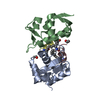
| ||||||||
|---|---|---|---|---|---|---|---|---|---|
| 1 |
| ||||||||
| Unit cell |
|
-Components
| #1: Protein | Mass: 13535.410 Da / Num. of mol.: 2 / Fragment: P53 BINDING DOMAIN, RESIDUES 6-125 / Mutation: YES Source method: isolated from a genetically manipulated source Source: (gene. exp.) Homo sapiens (human) / Gene: MDM2 / Plasmid: PGEX-6-P-1(MDM2-6-125) / Production host:  ESCHERICHIA COLI (E. coli) / Strain (production host): BL21(DE3) ESCHERICHIA COLI (E. coli) / Strain (production host): BL21(DE3)References: UniProt: Q00987, RING-type E3 ubiquitin transferase #2: Protein/peptide | Mass: 1541.850 Da / Num. of mol.: 2 / Fragment: MDM2 INTERACTING PEPTIDE, RESIDUES 17-27 / Source method: obtained synthetically / Source: (synth.) SYNTHETIC CONSTRUCT (others) #3: Water | ChemComp-HOH / | Water Mass: 18.015 Da / Num. of mol.: 85 / Source method: isolated from a natural source / Formula: H2O Mass: 18.015 Da / Num. of mol.: 85 / Source method: isolated from a natural source / Formula: H2ONonpolymer details | METHYLAMIN | |
|---|
-Experimental details
-Experiment
| Experiment | Method: X-RAY DIFFRACTION / Number of used crystals: 1 |
|---|
- Sample preparation
| Crystal | Density Matthews: 2.42 Å3/Da / Density % sol: 49.24 % / Description: NONE |
|---|---|
| Crystal grow | Temperature: 289 K / Method: vapor diffusion, sitting drop / pH: 6.4 Details: 0.04 M CITRIC ACID, 0.06 M BIS-TRIS PROPANE PH 6.4, AND 20% PEG 3350 |
-Data collection
| Diffraction | Mean temperature: 100 K |
|---|---|
| Diffraction source | Source: SYNCHROTRON / Site: NSRRC  / Type: NSRRC / Wavelength: 1 / Type: NSRRC / Wavelength: 1 |
| Detector | Date: Apr 7, 2014 |
| Radiation | Protocol: SINGLE WAVELENGTH / Monochromatic (M) / Laue (L): M / Scattering type: x-ray |
| Radiation wavelength | Wavelength: 1 Å / Relative weight: 1 |
| Reflection | Resolution: 1.97→30 Å / Num. obs: 19576 / % possible obs: 99.3 % / Observed criterion σ(I): 2 / Redundancy: 6.4 % / Rmerge(I) obs: 0.05 / Net I/σ(I): 34.74 |
| Reflection shell | Resolution: 1.97→2.02 Å / Redundancy: 5.8 % / Rmerge(I) obs: 0.34 / Mean I/σ(I) obs: 4.87 / % possible all: 95.3 |
- Processing
| Software |
| ||||||||||||||||||||||||||||||||||||||||||||||||||||||||||||||||||||||||||||||||||||||||||||||||||||||||||||||||||||||||||||||||||||||||||||||||||||||||||||||||||||||||||||||||||||||
|---|---|---|---|---|---|---|---|---|---|---|---|---|---|---|---|---|---|---|---|---|---|---|---|---|---|---|---|---|---|---|---|---|---|---|---|---|---|---|---|---|---|---|---|---|---|---|---|---|---|---|---|---|---|---|---|---|---|---|---|---|---|---|---|---|---|---|---|---|---|---|---|---|---|---|---|---|---|---|---|---|---|---|---|---|---|---|---|---|---|---|---|---|---|---|---|---|---|---|---|---|---|---|---|---|---|---|---|---|---|---|---|---|---|---|---|---|---|---|---|---|---|---|---|---|---|---|---|---|---|---|---|---|---|---|---|---|---|---|---|---|---|---|---|---|---|---|---|---|---|---|---|---|---|---|---|---|---|---|---|---|---|---|---|---|---|---|---|---|---|---|---|---|---|---|---|---|---|---|---|---|---|---|---|
| Refinement | Method to determine structure: MOLECULAR REPLACEMENT Starting model: PDB ENTRY 2AXI Resolution: 1.99→105.68 Å / Cor.coef. Fo:Fc: 0.954 / Cor.coef. Fo:Fc free: 0.926 / SU B: 8.541 / SU ML: 0.118 / Cross valid method: THROUGHOUT / ESU R: 0.169 / ESU R Free: 0.153 / Stereochemistry target values: MAXIMUM LIKELIHOOD Details: HYDROGENS HAVE BEEN ADDED IN THE RIDING POSITIONS. U VALUES WITH TLS ADDED
| ||||||||||||||||||||||||||||||||||||||||||||||||||||||||||||||||||||||||||||||||||||||||||||||||||||||||||||||||||||||||||||||||||||||||||||||||||||||||||||||||||||||||||||||||||||||
| Solvent computation | Ion probe radii: 0.8 Å / Shrinkage radii: 0.8 Å / VDW probe radii: 1.2 Å / Solvent model: MASK | ||||||||||||||||||||||||||||||||||||||||||||||||||||||||||||||||||||||||||||||||||||||||||||||||||||||||||||||||||||||||||||||||||||||||||||||||||||||||||||||||||||||||||||||||||||||
| Displacement parameters | Biso mean: 34.247 Å2
| ||||||||||||||||||||||||||||||||||||||||||||||||||||||||||||||||||||||||||||||||||||||||||||||||||||||||||||||||||||||||||||||||||||||||||||||||||||||||||||||||||||||||||||||||||||||
| Refinement step | Cycle: LAST / Resolution: 1.99→105.68 Å
| ||||||||||||||||||||||||||||||||||||||||||||||||||||||||||||||||||||||||||||||||||||||||||||||||||||||||||||||||||||||||||||||||||||||||||||||||||||||||||||||||||||||||||||||||||||||
| Refine LS restraints |
|