 Movie
Movie Controller

[English] 日本語
 Yorodumi
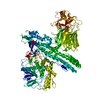
- PDB-4umk: The complex of Spo0J and parS DNA in chromosomal partition system -
+ Open data

- Basic information
| Entry | Database: PDB / ID: 4umk | ||||||
|---|---|---|---|---|---|---|---|
| Title | The complex of Spo0J and parS DNA in chromosomal partition system | ||||||
 Components Components |
| ||||||
 Keywords Keywords | DNA BINDING PROTEIN/DNA / DNA BINDING PROTEIN-DNA COMPLEX | ||||||
| Function / homology |  Function and homology information Function and homology informationpositive regulation of sporulation resulting in formation of a cellular spore /  chromosome segregation / chromosome / DNA binding chromosome segregation / chromosome / DNA bindingSimilarity search - Function | ||||||
| Biological species |   HELICOBACTER PYLORI (bacteria)BACILLUS SUBTILIS (bacteria) HELICOBACTER PYLORI (bacteria)BACILLUS SUBTILIS (bacteria) | ||||||
| Method | X-RAY DIFFRACTION / SYNCHROTRON / MAD / Resolution: 3.096 Å | ||||||
 Authors Authors | Chen, B.W. / Chu, C.H. / Tung, J.Y. / Hsu, C.E. / Hsiao, C.D. / Sun, Y.J. | ||||||
 Citation Citation | Journal: Proc. Natl. Acad. Sci. U.S.A. / Year: 2015 Title: Insights into ParB spreading from the complex structure of Spo0J and parS. Authors: Chen, B.W. / Lin, M.H. / Chu, C.H. / Hsu, C.E. / Sun, Y.J. | ||||||
| History |
|
- Structure visualization
| Structure viewer | Molecule: MolmilJmol/JSmol |
|---|
- Downloads & links
-Download
| PDBx/mmCIF format | 4umk.cif.gz | 410 KB | Display | PDBx/mmCIF format |
|---|---|---|---|---|
| PDB format | pdb4umk.ent.gz | 330.1 KB | Display | PDB format |
| PDBx/mmJSON format | 4umk.json.gz | Tree view | PDBx/mmJSON format | |
| Others |  Other downloads Other downloads |
-Validation report
| Arichive directory | https://data.pdbj.org/pub/pdb/validation_reports/um/4umkftp://data.pdbj.org/pub/pdb/validation_reports/um/4umk | HTTPS FTP |
|---|
-Related structure data
| Similar structure data |
|---|


-Links
 PDBj
PDBj


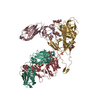
- Assembly
| Deposited unit | 
| ||||||||
|---|---|---|---|---|---|---|---|---|---|
| 1 | 
| ||||||||
| 2 | 
| ||||||||
| Unit cell |
|
-Components
| #1: Protein | Mass: 27382.582 Da / Num. of mol.: 4 Source method: isolated from a genetically manipulated source Source: (gene. exp.) HELICOBACTER PYLORI (bacteria) / Strain: 26695 / Production host: ESCHERICHIA COLI (E. coli) / Strain (production host): SG13009 / References: UniProt: O25758#2: DNA chain | Mass: 7483.842 Da / Num. of mol.: 2 / Source method: obtained synthetically / Source: (synth.) BACILLUS SUBTILIS (bacteria)#3: DNA chain | Mass: 7256.679 Da / Num. of mol.: 2 / Source method: obtained synthetically / Source: (synth.) BACILLUS SUBTILIS (bacteria)#4: Chemical | ChemComp-SO4 / Sulfate  Mass: 96.063 Da / Num. of mol.: 6 / Source method: obtained synthetically / Formula: SO4 Mass: 96.063 Da / Num. of mol.: 6 / Source method: obtained synthetically / Formula: SO4#5: Water | ChemComp-HOH / | Water Mass: 18.015 Da / Num. of mol.: 231 / Source method: isolated from a natural source / Formula: H2O Mass: 18.015 Da / Num. of mol.: 231 / Source method: isolated from a natural source / Formula: H2O |
|---|
-Experimental details
-Experiment
| Experiment | Method: X-RAY DIFFRACTION |
|---|
- Sample preparation
| Crystal | Density Matthews: 2.53 Å3/Da / Density % sol: 51 % / Description: NONE |
|---|---|
| Crystal grow | pH: 8 / Details: PH 8 |
-Data collection
| Diffraction | Mean temperature: 293 K |
|---|---|
| Diffraction source | Source: SYNCHROTRON / Site: NSRRC  / Beamline: BL13B1 / Wavelength: 1 / Beamline: BL13B1 / Wavelength: 1 |
| Detector | Type: ADSC CCD / Detector: CCD |
| Radiation | Protocol: MAD / Monochromatic (M) / Laue (L): M / Scattering type: x-ray |
| Radiation wavelength | Wavelength: 1 Å / Relative weight: 1 |
| Reflection | Resolution: 3.1→30 Å / Num. obs: 33332 / % possible obs: 99.8 % / Observed criterion σ(I): 3 / Redundancy: 4.2 % / Biso Wilson estimate: 73.03 Å2 / Rmerge(I) obs: 0.07 / Net I/σ(I): 17 |
| Reflection shell | Resolution: 3.1→3.21 Å / Redundancy: 4.3 % / Rmerge(I) obs: 0.44 / Mean I/σ(I) obs: 3.1 / % possible all: 100 |
- Processing
| Software |
| |||||||||||||||||||||||||||||||||||||||||||||||||||||||||||||||||||||||||||||||||||||||||||||||||||||||||
|---|---|---|---|---|---|---|---|---|---|---|---|---|---|---|---|---|---|---|---|---|---|---|---|---|---|---|---|---|---|---|---|---|---|---|---|---|---|---|---|---|---|---|---|---|---|---|---|---|---|---|---|---|---|---|---|---|---|---|---|---|---|---|---|---|---|---|---|---|---|---|---|---|---|---|---|---|---|---|---|---|---|---|---|---|---|---|---|---|---|---|---|---|---|---|---|---|---|---|---|---|---|---|---|---|---|---|
| Refinement | Method to determine structure: MAD Starting model: NONE Resolution: 3.096→23.574 Å / SU ML: 0.47 / σ(F): 0 / Phase error: 35.19 / Stereochemistry target values: ML Details: CHAIN A RESIDUES 1-54, 227-240 CHAIN B RESIDUES 1-44, 228-240 CHAIN C RESIDUES 1-34, 228-240 CHAIN D RESIDUES 1-50, 227-240 ARE MISSING.
| |||||||||||||||||||||||||||||||||||||||||||||||||||||||||||||||||||||||||||||||||||||||||||||||||||||||||
| Solvent computation | Shrinkage radii: 1.1 Å / VDW probe radii: 1.2 Å / Solvent model: FLAT BULK SOLVENT MODEL | |||||||||||||||||||||||||||||||||||||||||||||||||||||||||||||||||||||||||||||||||||||||||||||||||||||||||
| Displacement parameters | Biso mean: 67.7 Å2 | |||||||||||||||||||||||||||||||||||||||||||||||||||||||||||||||||||||||||||||||||||||||||||||||||||||||||
| Refinement step | Cycle: LAST / Resolution: 3.096→23.574 Å
| |||||||||||||||||||||||||||||||||||||||||||||||||||||||||||||||||||||||||||||||||||||||||||||||||||||||||
| Refine LS restraints |
| |||||||||||||||||||||||||||||||||||||||||||||||||||||||||||||||||||||||||||||||||||||||||||||||||||||||||
| LS refinement shell |
| |||||||||||||||||||||||||||||||||||||||||||||||||||||||||||||||||||||||||||||||||||||||||||||||||||||||||
| Refinement TLS params. | Method: refined / Origin x: 59.2522 Å / Origin y: 26.798 Å / Origin z: 34.8308 Å
| |||||||||||||||||||||||||||||||||||||||||||||||||||||||||||||||||||||||||||||||||||||||||||||||||||||||||
| Refinement TLS group | Selection details: ALL |
