 Movie
Movie Controller

[English] 日本語
 Yorodumi
- PDB-4ufr: Structure of the ectodomain of LGR5 in complex with R-spondin-2 (... -
+ Open data

- Basic information
| Entry | Database: PDB / ID: 4ufr | ||||||
|---|---|---|---|---|---|---|---|
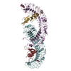
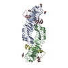
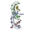
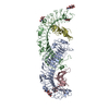
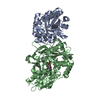
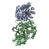
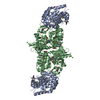
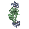
| Title | Structure of the ectodomain of LGR5 in complex with R-spondin-2 (Fu1Fu2) | ||||||
 Components Components |
| ||||||
 Keywords Keywords |  SIGNALING PROTEIN / WNT / LGR / RSPO SIGNALING PROTEIN / WNT / LGR / RSPO | ||||||
| Function / homology |  Function and homology information Function and homology informationtrachea cartilage morphogenesis / negative regulation of odontogenesis of dentin-containing tooth / lung growth / epithelial cell proliferation involved in renal tubule morphogenesis / oocyte differentiation / protein-hormone receptor activity / Regulation of FZD by ubiquitination / dopaminergic neuron differentiation / G protein-coupled peptide receptor activity / embryonic forelimb morphogenesis ...trachea cartilage morphogenesis / negative regulation of odontogenesis of dentin-containing tooth / lung growth / epithelial cell proliferation involved in renal tubule morphogenesis / oocyte differentiation / protein-hormone receptor activity / Regulation of FZD by ubiquitination / dopaminergic neuron differentiation / G protein-coupled peptide receptor activity / embryonic forelimb morphogenesis / embryonic hindlimb morphogenesis / epithelial tube branching involved in lung morphogenesis / bone mineralization / inner ear development / hair follicle development / canonical Wnt signaling pathway / Regulation of FZD by ubiquitination / trans-Golgi network membrane / G protein-coupled receptor activity / adenylate cyclase-activating G protein-coupled receptor signaling pathway / osteoblast differentiation / transmembrane signaling receptor activity / positive regulation of canonical Wnt signaling pathway / heparin binding / regulation of cell population proliferation / G protein-coupled receptor signaling pathway / signaling receptor binding / Golgi apparatus / cell surface / extracellular region / nucleoplasm / plasma membraneSimilarity search - Function | ||||||
| Biological species |  HOMO SAPIENS (human) HOMO SAPIENS (human) | ||||||
| Method | X-RAY DIFFRACTION / SYNCHROTRON / OTHER / Resolution: 2.2 Å | ||||||
 Authors Authors | Zebisch, M. / Jones, E.Y. | ||||||
 Citation Citation | Journal: J.Struct.Biol. / Year: 2015 Title: Crystal Structure of R-Spondin 2 in Complex with the Ectodomains of its Receptors Lgr5 and Znrf3. Authors: Zebisch, M. / Jones, E.Y. | ||||||
| History |
|
- Structure visualization
| Structure viewer | Molecule: MolmilJmol/JSmol |
|---|
- Downloads & links
-Download
| PDBx/mmCIF format | 4ufr.cif.gz | 449.3 KB | Display | PDBx/mmCIF format |
|---|---|---|---|---|
| PDB format | pdb4ufr.ent.gz | 375.4 KB | Display | PDB format |
| PDBx/mmJSON format | 4ufr.json.gz | Tree view | PDBx/mmJSON format | |
| Others |  Other downloads Other downloads |
-Validation report
| Arichive directory | https://data.pdbj.org/pub/pdb/validation_reports/uf/4ufrftp://data.pdbj.org/pub/pdb/validation_reports/uf/4ufr | HTTPS FTP |
|---|
-Related structure data


-Links
 PDBj
PDBj


- Assembly
| Deposited unit | 
| ||||||||
|---|---|---|---|---|---|---|---|---|---|
| 1 |
| ||||||||
| Unit cell |
|
-Components
| #1: Protein | Mass: 54052.348 Da / Num. of mol.: 2 / Fragment: ECTODOMAIN, RESIDUES 32-487 AND RESIDUES 538-544 Source method: isolated from a genetically manipulated source Details: UNSTRUCTURED LOOP REPLACED WITH SHORT LINKER, A488-H537 TO NGNNGD Source: (gene. exp.) HOMO SAPIENS (human) / Plasmid: PHLSEC / Cell line (production host): HEK293T / Production host: HOMO SAPIENS (human) / References: UniProt: O75473#2: Protein | Mass: 13946.869 Da / Num. of mol.: 2 / Fragment: FU1-FU2, RESIDUES 39-144 Source method: isolated from a genetically manipulated source Source: (gene. exp.) HOMO SAPIENS (human) / Plasmid: PHLSEC / Cell line (production host): HEK293T / Production host: HOMO SAPIENS (human) / References: UniProt: Q8BFU0#3: Sugar | N-Acetylglucosamine  Type: D-saccharide, beta linking / Mass: 221.208 Da / Num. of mol.: 2 Type: D-saccharide, beta linking / Mass: 221.208 Da / Num. of mol.: 2Source method: isolated from a genetically manipulated source Formula: C8H15NO6 #4: Chemical | ChemComp-CL / Chloride  Mass: 35.453 Da / Num. of mol.: 6 / Source method: obtained synthetically / Formula: Cl Mass: 35.453 Da / Num. of mol.: 6 / Source method: obtained synthetically / Formula: Cl#5: Water | ChemComp-HOH / | Water Mass: 18.015 Da / Num. of mol.: 150 / Source method: isolated from a natural source / Formula: H2O Mass: 18.015 Da / Num. of mol.: 150 / Source method: isolated from a natural source / Formula: H2O |
|---|
-Experimental details
-Experiment
| Experiment | Method: X-RAY DIFFRACTION / Number of used crystals: 3 |
|---|
- Sample preparation
| Crystal | Density Matthews: 2.5 Å3/Da / Density % sol: 50 % / Description: NONE |
|---|---|
| Crystal grow | pH: 5.5 Details: 25 %W/V PEG3350, 0.200 M LITHIUM SULPHATE, 0.100 M BIS-TRIS PH 5.5 |
-Data collection
| Diffraction | Mean temperature: 100 K |
|---|---|
| Diffraction source | Source: SYNCHROTRON / Site: Diamond  / Beamline: I04 / Wavelength: 1.009 / Beamline: I04 / Wavelength: 1.009 |
| Detector | Type: DECTRIS PILATUS / Detector: PIXEL / Date: Oct 25, 2013 |
| Radiation | Protocol: SINGLE WAVELENGTH / Monochromatic (M) / Laue (L): M / Scattering type: x-ray |
| Radiation wavelength | Wavelength: 1.009 Å / Relative weight: 1 |
| Reflection | Resolution: 2.2→69 Å / Num. obs: 62167 / % possible obs: 90.6 % / Observed criterion σ(I): -3 / Redundancy: 7.6 % / Biso Wilson estimate: 37 Å2 / Rmerge(I) obs: 0.12 / Net I/σ(I): 10.8 |
- Processing
| Software | Name: REFMAC / Version: 5.8.0073 / Classification: refinement | ||||||||||||||||||||||||||||||||||||||||||||||||||||||||||||||||||||||||||||||||||||||||||||||||||||||||||||||||||||||||||||||||||||||||||||||||||||||||||||||||||||||||||||||||||||||
|---|---|---|---|---|---|---|---|---|---|---|---|---|---|---|---|---|---|---|---|---|---|---|---|---|---|---|---|---|---|---|---|---|---|---|---|---|---|---|---|---|---|---|---|---|---|---|---|---|---|---|---|---|---|---|---|---|---|---|---|---|---|---|---|---|---|---|---|---|---|---|---|---|---|---|---|---|---|---|---|---|---|---|---|---|---|---|---|---|---|---|---|---|---|---|---|---|---|---|---|---|---|---|---|---|---|---|---|---|---|---|---|---|---|---|---|---|---|---|---|---|---|---|---|---|---|---|---|---|---|---|---|---|---|---|---|---|---|---|---|---|---|---|---|---|---|---|---|---|---|---|---|---|---|---|---|---|---|---|---|---|---|---|---|---|---|---|---|---|---|---|---|---|---|---|---|---|---|---|---|---|---|---|---|
| Refinement | Method to determine structure: OTHER Starting model: NONE Resolution: 2.2→110.67 Å / Cor.coef. Fo:Fc: 0.954 / Cor.coef. Fo:Fc free: 0.917 / SU B: 23.344 / SU ML: 0.254 / Cross valid method: THROUGHOUT / ESU R: 0.283 / ESU R Free: 0.229 / Stereochemistry target values: MAXIMUM LIKELIHOOD / Details: HYDROGENS HAVE BEEN ADDED IN THE RIDING POSITIONS.
| ||||||||||||||||||||||||||||||||||||||||||||||||||||||||||||||||||||||||||||||||||||||||||||||||||||||||||||||||||||||||||||||||||||||||||||||||||||||||||||||||||||||||||||||||||||||
| Solvent computation | Ion probe radii: 0.8 Å / Shrinkage radii: 0.8 Å / VDW probe radii: 1.2 Å / Solvent model: MASK | ||||||||||||||||||||||||||||||||||||||||||||||||||||||||||||||||||||||||||||||||||||||||||||||||||||||||||||||||||||||||||||||||||||||||||||||||||||||||||||||||||||||||||||||||||||||
| Displacement parameters | Biso mean: 59.664 Å2
| ||||||||||||||||||||||||||||||||||||||||||||||||||||||||||||||||||||||||||||||||||||||||||||||||||||||||||||||||||||||||||||||||||||||||||||||||||||||||||||||||||||||||||||||||||||||
| Refinement step | Cycle: LAST / Resolution: 2.2→110.67 Å
| ||||||||||||||||||||||||||||||||||||||||||||||||||||||||||||||||||||||||||||||||||||||||||||||||||||||||||||||||||||||||||||||||||||||||||||||||||||||||||||||||||||||||||||||||||||||
| Refine LS restraints |
|